
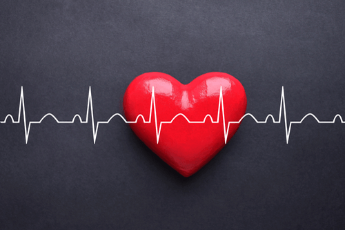
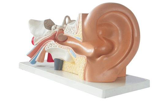
Echocardiography – ECHO

Last Updated December 20th, 2021
What is echocardiography?
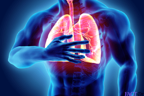
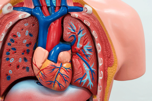
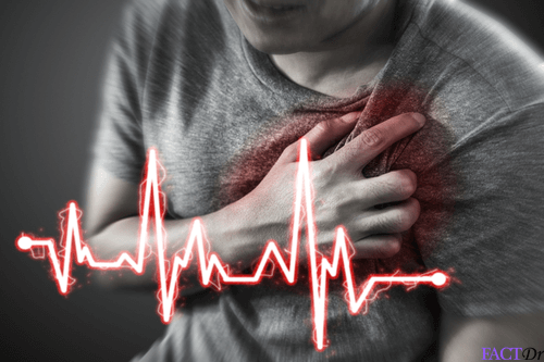
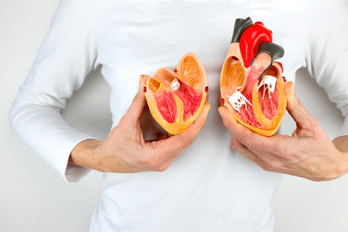
Echocardiography (commonly known as ECHO) is an imaging modality (similar to ultrasonography) used to visualize the interior of your heart. An echocardiogram employs special sound waves to produce graphic images of the heart.
What are the indications for echocardiography?
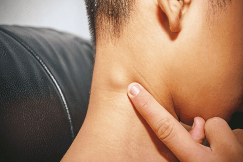
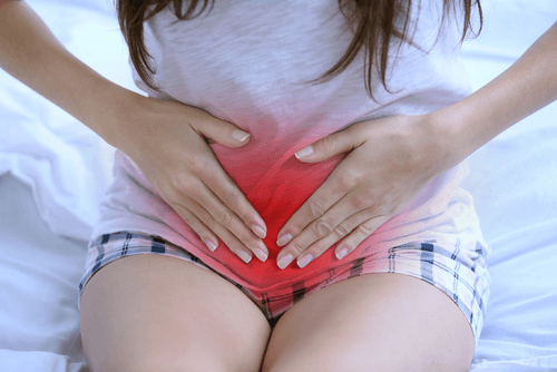
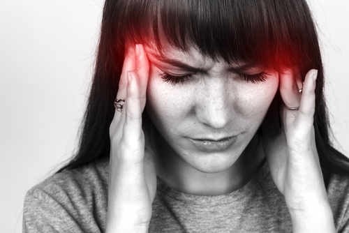
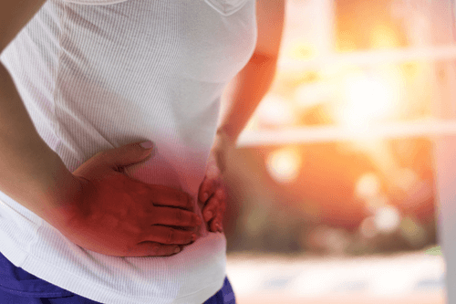
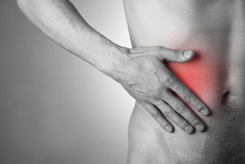
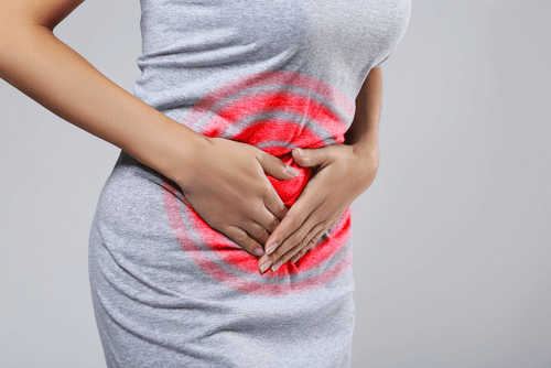
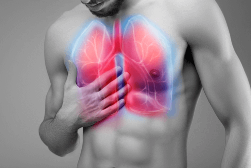
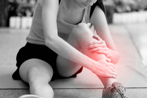
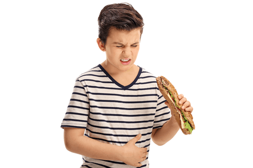
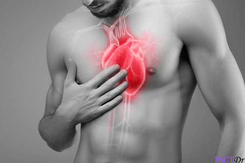
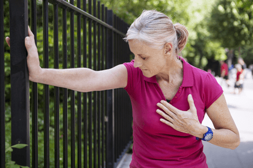
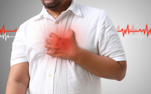
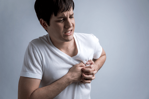
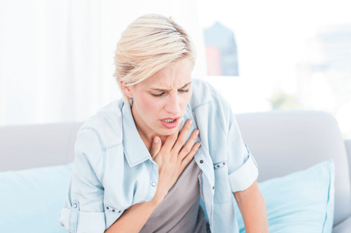
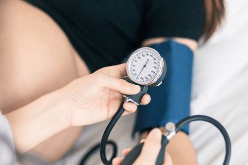
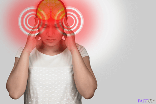
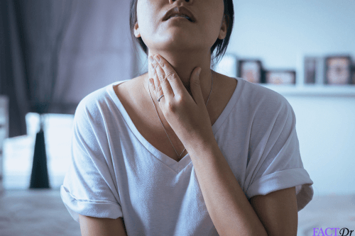
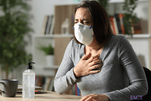
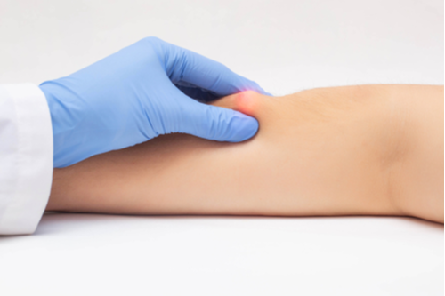
Your physician may order echocardiography for you if you present with features of heart disease. These include- shortness of breath, swelling in the legs, palpitations, dizziness and chest pain. Echocardiography is also indicated in infants and children in order to detect congenital heart defects.
What can an echocardiography test detect?
An echocardiography test can detect changes in-
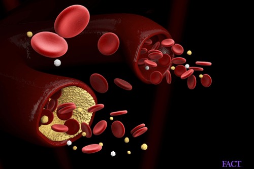
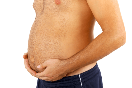
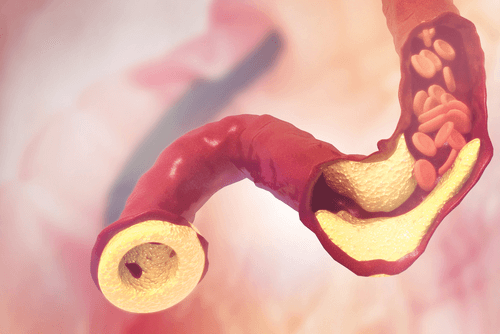
The size of heart (Cardiomegaly)- The size of heart increases in conditions such as hypertension (known as hypertrophic cardiomyopathy, it involves the thickening of the walls of the left lower chamber of the heart-the left ventricle). The heart can also enlarge in size secondary to alcoholism, diabetes, and coronary artery disease. In these cases, the disorder is known as dilated cardiomyopathy.
The shape of heart- Apart from the conditions described above, the heart can change shape with advancing age, in tetralogy of Fallot (a congenital disease) and Takotsubocardiomyopathy (a stress-induced disorder).
The functioning of your heart– Any compromise with the blood flow to the heart (either from a previous heart attack or an ongoing cardiac insult) can weaken the cardiac muscles. These weak muscles are unable to pump blood effectively.
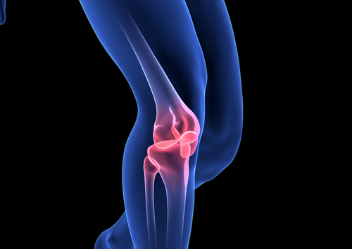
The functioning of the heart valves– An echocardiogram can detect if the valves of the heart are opening and closing efficiently. Any stenosis or regurgitation of the valves can be visualized.
Structural imaging of the great vessels– The vessels visualized include the aorta and the pulmonary vessels.
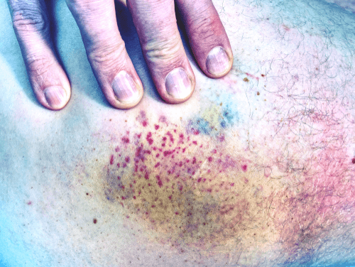
Structural imaging of the pericardium (the covering of the heart)-Diseases such as pericardial effusion and infective pericarditis can be detected.
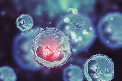
Congenital diseases– An echocardiography test can detect congenital cardiac diseases in a fetus (fetal echocardiogram).
What are the different types of echocardiography tests?
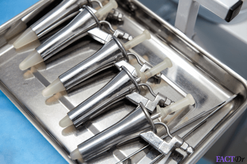
Transthoracic echocardiography (commonly referred to as TTE) is the most common variety of echocardiography tests. It uses a device called a transducer to send sound waves to your heart via the chest wall. As these waves are reflected by the heart, a computer converts them into images that can be viewed on a screen. In this article, we will be talking about TTE mostly.
Stress echocardiography– It is a type of echocardiography where the heart is made to beat faster and harder by exposing it to stress. The subsequent procedure is the same as TTE. The stress may be in the form of physical activity (running on a treadmill or cycling a stationary bike) or drug-induced.
This technique helps in unmasking diseases of the heart which may not be visible during normal echocardiography (e.g. coronary artery disease).
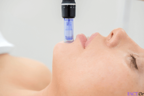
Transesophageal Echocardiography (TEE)– To visualize the interior of your heart with more clarity, the physician may attach the transducer to the end of a flexible tube and insert it in your throat and into your esophagus (food pipe).
As the ultrasound waves have a lesser distance to travel, the images obtained are of very high quality. Three-dimensional echocardiography- 3-D images with a very high resolution are produced.
Fetal echocardiography– It is performed between 18-22 weeks of pregnancy in order to look for any congenital cardiac defect in the fetus. The transducer is moved over the abdominal region of the pregnant female and the images procured.
Doppler echocardiography– It employs the ‘Doppler phenomenon’ to ascertain the speed and direction of blood flow in the heart and its major blood vessels. It does so by detecting changes in the frequency of the ‘backscatter signal’ from small moving structures.
Can I undergo echocardiography if I am pregnant?
An echocardiography test does not use any radiation. Therefore, it is perfectly safe to undergo echocardiography during pregnancy. In fact, fetal echocardiography is performed to detect congenital heart diseases before birth.
Can children undergo echocardiography test?
Yes. It is safe for children to undergo echocardiography. No radiation is used during this process.
What are the risks involved?
Echocardiography is a non-invasive, painless and safe procedure.
– A dye is used sometimes used in echocardiography in order to visualize the interior of the heart better. An allergic reaction to the dye is rare but at times a serious complication.
– During transesophageal echocardiography you may feel pain, bruising, bleeding and subsequently infection at the site of intravenous access. You may experience soreness in your throat for some time. A sedative is used in TEE in many cases. You may experience side effects related to it.esophageal perforation–1 in 10,000 is another rare complication.
– During stress echocardiography, there is a risk of precipitating an underlying heart disorder. But these risks are related to the stress-inducing exercise/medicine and not directly linked with the procedure.
What are the contra-indications of echocardiography?
There are no absolute contra-indications for echocardiography. However, as per the FDA (US Food and Drug Administration) guidelines, transthoracic echo test should not be performed in the following cases-
– Acute MI (myocardial infarction) or acute coronary syndrome
– Heart failure (worsening or unstable)
– Serious ventricular arrhythmia
– Respiratory failure
– Conditions that may lead to pulmonary hypertension
How should I prepare for echocardiography?
There are no specific precautions required on your part before undergoing am echocardiography test. However, if you are undergoing a TEE, you need to fast for 6-8 hours prior to the test. Moreover, you need to talk to your physician regarding your medical history, past drug history, any medicines that you might be taking and allergies that you suffer from.
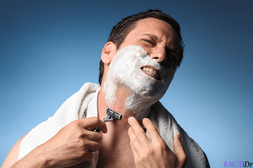
It is advisable to remove any precious jewelry before coming to the hospital for the test. Remove any piercings on your chest as these may interfere with the transducer. Males undergoing this procedure are sometimes advised to shave off their chest hair to make it easier for the electrodes to be patched onto the chest and facilitate their removal at the end of the procedure.
Do I need to fast before an echocardiography exam?
There is no need to fast before an echocardiography test. However, if you are supposed to undergo TEE, you need to fast for 6-8 hours prior to the test.
How is an echocardiography test performed?
An echocardiogram is performed on an outpatient basis- either in a hospital or a diagnostic center.
What happens before the echocardiography procedure begins?
Before the procedure-
– Your physician will explain the procedure of echocardiography to you.
– You will be asked to remove your clothes, waist up. If you are a female, you will be given a hospital gown to wear.
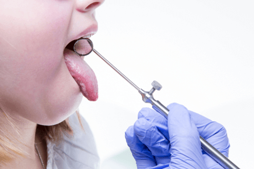
How is an echocardiography procedure performed?
– You will be made to lie on an examination table on your side (the left lateral decubitus position is commonly used). You will be asked to place your left arm over your head.
– You may be asked to change positions during the procedure and to hold your breath at times.
– Electrodes (usually 3 in number) will be applied to your chest. These are soft, sticky flat patches that help in recording the electrical activity of your heart in the form of an ECG (electrocardiogram).
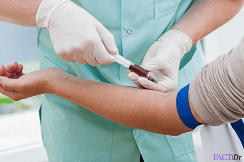
– A wand-like an instrument known as the ultrasound transducer will be moved over the area of your chest. A gel is applied both on the wand and your chest to increase the conductibility of the waves.
– You may feel some discomfort when the transducer is pressed firmly against your chest.
– The transducer performs the function of sending ultrasonic waves to your heart.
-As your heart sends echoes of the sound waves, these are detected by the transducer and conveyed to a computer. The computer converts these waves into images that can be stored in the form of discs.
What happens after the procedure is over?
After the procedure-
– There are no specific instructions to be followed once the test is over.
– If you have undergone a transesophageal echo, you will be shifted to a recovery room to recover from the effects of the sedative used.
How long does an echocardiography exam take?
A standard echocardiography test takes about 45-60 minutes to be conducted.
What is the cost of undergoing echocardiography in India?
The cost of undergoing echocardiography in India ranges from INR 1500-5000.
What are the advantages and limitations of an echocardiography test?
| Advantages | Limitations |
| It is a non-invasive, painless and safe procedure. | In patients who are obese, it is difficult to perform echocardiography as the fat may impede the transmission of sound waves. |
| No radiation is employed. | In very thin patients, the images may be hard to produce due to overcrowding by the ribs. |
| Safe to be used in pregnant and lactating females. | The technical acumen of the sonographer determines the quality of the images. |
| Can be used to detect anomalies in a fetus. | It is a costly procedure. |
Subscribe to free FactDr newsletters.
REVAMP YOUR
LIFE
HEALTH
WELLNESS
If you're enjoying our website, we promise you'll absolutely love our new posts. Be the first one to get a copy!
Get factually correct, actionable tips delivered straight to your inbox once a week.
We hate spam too. We will never share your email address with anyone. If you change your mind later, you can unsubscribe with just one click
By clicking Subscribe, I agree to the FactDr Terms & Conditions & Privacy Policy and understand that I may opt out of FactDr subscriptions at any time.
Help Others Be Fit
Top Stories
- Blue Balls

- Ferritin Test
- Rubella IgG Test
- Tonsil Stones
- Enterogermina

- Muteness

- Random Blood Sugar Test
- Sebaceous Cyst

- Lectin: The common link of proteins between peas and people

- Augmentin 625

- Lipid Profile
- Primolut N
- Bifilac

- Cervical Cysts
- Anti-Thyroglobulin Antibody test

- Pulmonary Function Tests
- Lupus Rash
- Pott’s disease

- Dexorange

- FBS Test – Fasting Blood Sugar
- EKG – Electrocardiogram

- Rheumatoid Factor (RF)

- Flunarizine
- Tongue infections
- Leukoplakia
- The real reason why you shouldn’t be eating ramen noodles

- Skin Rashes

- Betadine
- TBHQ: A carcinogen lingering in your child’s favorite snacks

- Post-Prandial Blood Sugar
- T Bact Ointment

- Thyroglobulin (TG) Test
- Dyshidrotic eczema

- Serum Electrolyte
- Becosules

- Passion fruit: How one exotic fruit can help you fight infections and cancer!
- Hydrocele

- Beriberi

- Orofer XT

- Chondromalacia
- Thyromegaly

- Cervical Polyps

- Amoebiasis

- Blue Waffle Disease
- High platelet count

- Hepatitis B Envelope Antigen (HBeAg)

- Avil

- Treponema Pallidum Antibody(TPAB) test

- Arterial Blood Gas Analysis

- Nurokind LC
- Inverted nipples
- AST- Aspartate Aminotransferase Test
- Serum Zinc Test

- Herpes Simplex Virus I (HSV)-IgG Test

- Chiggers

- The importance of roughage in diet

- Evion LC

- Hangnails

- Duphalac
- Cherry Angioma

- Diverticulitis diet: The right way to eat if you suffer from the disease

- Wasp Sting

- HLA-B27 test
- Myospaz

- Balanitis

- Non-Hodgkin’s Lymphoma
- Free Triiodothyronine (FT3) Test
- Abscess
- DHEA Sulfate (DHEAS) Test
- Chromium Toxicity

- Hypermetropia
- Betnovate
- IgE test – Immunoglobulin E

- Pus
- Abdominal CT scan
- Pellagra
- Metabolic Disorders

- Yellow poop
- Autoimmune Diseases: Find out if your body is attacking you right now

- Cardiolipin Antibody (ACL) –IgM Test

- Dengue NS1 test

- Beta 2 Glycoprotein 1 IgG
- Vitamin K2: 8 reasons why you need this bone-building & cancer-fighting nutrient

- Fibroadenoma

- Prickly Heat Rash
- Silicon Dioxide: How can a component of sand be essential to your wellbeing?

- Maltitol: Things you must know about this artificial sweetener
- Albinism

- Enlarged Liver
- Racecadotril

- Viral Infections
- Zerodol

- Urinary Microalbumin Test
- Helicobacter Pylori – IgG Test
- Erythropoietin (EPO) Test
- Endomysial Antibodies (EMA) Test

- Fatty Liver Disease
- Brown Recluse Spider Bites
- Taxim O
- Shelcal 500

- Ketorol DT

- Kidney Cysts
- The Big 5 lifestyle diseases: How your everyday living might be killing you

- Excretory System Diseases

- Smallpox

- Gum Disease
- Clavam 625

- Pan D

- Swollen Lymph Nodes
- Bilirubin Test
- Sickle cell disease
- Klinefelter’s Syndrome

- How do neutrophils protect you from fatal bacterial attacks?

- Chymoral Forte
- Brain Cysts

- Cavernous Sinus Thrombosis
- Intestinal Adhesions

- Smegma
- CLA: A breakthrough weight loss supplement with minimal side effects

- Birthmarks
- Cytomegalovirus (CMV)- IgG Test
- Eustachian Tube : Functions and top home remedies to prevent its infection

- Free thyroxine test (FT4)
- Chromium Picolinate: An essential mineral supplement for faster weight loss

- Alexandria’s Genesis
- Skin Tags

- Cellulitis
- Antinuclear Antibody Test – (ANA)
- Enteritis

- Genital Herpes

- Maltodextrin: What are the hidden health benefits of this food additive?

- Ingrown Toenail
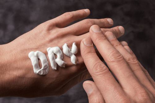
- Dry skin
- Cheilitis
- Phlebitis

- Poop chart: Top things you didn’t you your poop could reveal about you

- Occupational Hazards: How to vouch for your health at your workplace

- Mox 500
- Levosulpiride

- Anti-CCP Test – Anti-Cyclic Citrullinated Peptide
- Disodium Hydrogen Citrate
- Genital Warts (HPV)

- Myocardial infarction

- Benign Tumours
- Scabies
- Jock Itch
- Hydrocephalus

- Zerodol SP

- Prolapsed Uterus

- Duphaston
- BRAT Diet: What is the right way to follow this diarrhea-relieving diet?

- Tissue Transglutaminase Antibody (tTG) Test
- Internal Bleeding
- Hepatitis Profile
- Etizolam

- Deep Vein Thrombosis
- Burning mouth syndrome
- Zifi 200

- Anti Ds-DNA antibody Test
- Norflox TZ
- Abnormal Vaginal Bleeding
- Cystoscopy

- Neural tube defects

- Salivary Gland Infection
- Electroencephalogram – EEG

- Sputum test
- Rectal Prolapse

- Gonorrhoea

- Cardiac Profile
- Top 6 remedies to treat a razor burn at home
- Temper Tantrum

- Foot Corns

- Asthenia

- 17 OH Progesterone test
- Nexito Plus

- Pinworms
- Poison oak

- Pyelonephritis

- Stye
- Lisp

- Bronchitis
- Dysentery
- Mumps
- Moon Facies ( Cushing Syndrome)

- Eating Disorders: Lifestyle choice or a psychological condition?
- Scalp Psoriasis
- Dwarfism

- Typhus

- Folate test

- Hypersplenism

- Rubella

- Hiatal Hernia

- Skin irritation
- Endocrine System Disorders

- Gilbert’s Syndrome
- Razo D

- Ringworm
- Athlete’s Foot
- Blood blister

- Deafness
- Fungal infections

- Hives
- Swollen Feet
- Helicobacter Pylori – IgA Test
- Genetic diseases
- Hemolytic Anemia
- Tonsillitis

- Arachnoid Cysts
- Carrageenan: How a simple seaweed extract could better your gut health & immunity
- Amylase Test

- Cerebral Cavernoma

- Flagyl 400
- Anti-Microsomal Antibody AMA Test

- Immunoglobulin M Test

- Common diseases that could cripple your vital organs

- Emphysema
- Anencephaly

- Proctitis

- Androstenedione Test
- Drotin DS
- Oxycodone

- CBC (Hemogram 6-part diff) blood test

- Oral Glucose Tolerance Test – GTT
- Stye : The best natural home remedies to ease the pain

- Piaget stages: Do they accurately describe the way the human brain develops?

- Lipoma
- Heat Rash
- Costochondritis

- Trisodium Phosphate: How a paint thinner made its way into your breakfast

- The science behind daith piercing: Can it really cure migraine?

- Copper Serum Test

- Arm fracture

- West Nile Disease

- Leukocytosis
- Maladaptive Daydreaming

- Tonometry

- Troubled with IBS? Here’s a complete roadmap to the low FODMAP diet

- Ulcers

- Panera Bread: The truth behind this “healthy” restaurant chain

- Ciplox Tz
- MRSA – Methicillin-Resistant Staphylococcus Aureus

- Beta hCG Test

- Sepsis (Blood Poisoning)
- Cloudy Urine

- Hematuria
- Free PSA Test
- Blood Disorders

- 9 benefits of walking we bet you didn’t know!

- Stuffy Nose

- Sitophobia

- Squid Ink: A unique food coloring and flavoring agent
- Oral Leukoplakia
- 5 unbelievable effects of dance on your overall health!

- Hand Fracture

- Werner’s Syndrome

- Dislocated Jaw

- Acanthosis nigricans
- Syphilis

- How are BMI and BMR different and what do these numbers mean?

- Blood thinners
- Rickets
- Adenoiditis

- Sinarest

- Ascites

- 8 hidden causes of obesity you probably didn’t know!

- Giardiasis
- How can berberine supplements help you live longer and healthier?

- Freckles

- Spider Bites

- Comedones
- Enlarged Heart

- Albumin Test
- Varicocele

- Milk Thistle: Find possible cures for fatal diseases in these purple blooms
- Tongue cancer
- Caralluma Fimbriata: How to eat this cactus to lose weight

- Anemia Profile

- Trichomoniasis

- These are the top foods to increase your hemoglobin count
- GGT – Gamma-Glutamyl Transferase Test
- Hookworms
- Quadriplegia

- Wound debridement and dressing

- Don’t let your sleep deficit grow into memory loss or heart attack

- Oligohydramnios

- Pre-cancerous Skin Lesions

- Lipoprotein (A) Test
- Lockjaw
- CA 15-3 Test

- Baby Acne

- Atelectasis

- How accurate is the hair follicle drug test?
- Congenital Glaucoma

- Canker Sores (Apthous Stomatitis)
- Beta 2 Glycoprotein 1 IgM
- Rhabdomyolysis
- Yellow Jacket Sting

- The real reasons for your mood swings and how to overcome them

- Charley Horse

- Power up your gut: 8 proven steps on how to improve your digestion
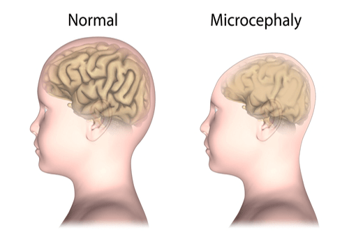
- Microcephaly
- Blisters
- Are you depressed or just stressed? Know when to see a doctor

- Addison’s Disease

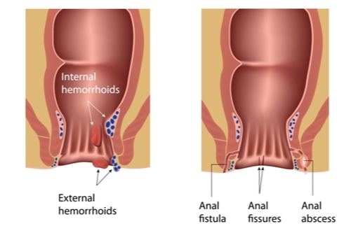
- Anal Abscess

- Motion Sickness

- Folliculitis

- Dyscalculia

- LP-PLA2 Test

- Tongue Diseases

- Ketoacidosis
- Plantar Warts
- Ingrown Hair
- Anhidrosis

- Edema

- Meftal Spas
- Sustan 200

- Chlorella: Therapeutic powers of the billion-year-old algae now within your reach
- Lipodystrophy
- Thalassemia
- Xylitol : Is this the right sugar substitute for you?
- Brain-Eating Amoeba (Naegleriasis)

- Signoflam
- Oxalates: How healthy greens can sometimes be bad news for your kidneys
- Staph Infection

- Green Healing: The amazing health benefits of being in nature

- Bruises
- What are Shiitake Mushrooms and why should you eat them?

- Cheston Cold
- Skin Problems
- Jicama: A fiber-rich tuber for your weight loss goals

- Bone tumor

- Silicosis
- Is bronchitis contagious? And how to prevent this infection?

- Insect Bites
- Chicken Pox

- Otitis media

- Blood Element Analysis Test
- Jackfruit: Slice your way into higher immunity, better digestion, & more…

- Joint Damage
- Carcinoembryonic antigen (CEA) test
- Appendicitis
- 7 liver detox facts and why ignoring these could kill you!

- Jaundice

- Green poop : What are the reasons and how can you correct it

- Hyperthyroidism
- Herpes Simplex Virus (HSV)-IgM Test
- Hypocalcemia
- Vitamin B12 Test
- Skull Fracture

- Astaxanthin: Why experts believe it could be 550x more potent than vitamin E!

- Itching

- Poison sumac
- Thyroid Scan

- Quinine: How gin and tonic came to be known as a potent health elixir

- Flat Feet

- Dengue Fever
- Itraconazole
- Pneumothorax
- Syncope

- Connective Tissue Diseases

- Colloid Cyst

- Molluscum contagiosum
- Skin Problems in Children

- Contact dermatitis
- Pneumonia
- Speech disorders

- Night Blindness

- Non-Allergic Rhinitis
- Lupus

- Subdural Hematoma
- Coconut sugar: What is it and is it healthier than table sugar?

- Nail Fungus
- CT – Computed Tomography Scan

- Iron Deficiency Profile
- Dragon Fruit – Blessing from Nature’s Basket

- Stages of wound healing

- Bulimia Nervosa

- Cold Sores
- Combiflam
- Quadriceps: Why stretching these is a must before going for a run

- Endometritis

- What to eat, what not to eat in the South Beach Diet?

- Bacterial Pneumonia
- Ear Infections
- 5 things you didn’t know about balneotherapy and how you can do it at home

- Scurvy

- Xanthan Gum: The pros and cons of this synthetic gummy additive

- Colon Polyps
- Tuberculosis
- Anemia

- Metal toxicity
- Arsenic Poisoning

- Surgery or home-remedy; what is the right way to treat a burn
- Hodgkin’s Disease

- A complete list of low-fat diets and how to follow them

- Lymphoma

- Trypophobia
- Hysteroscopy

- Fibrocystic Breast Changes
- Juice Plus: Can you really replace actual fruits with pills?

- Cystatin C Test
- Down syndrome

- Gastritis

- Stretch Marks
- Peripheral Artery Disease

- Pernicious anemia

- Pain disorder

- Yeast Infection
- Pressure ulcers

- Bell’s palsy

- Hymenoplasty

- The actual science behind smelling salts and how it helps in injury recovery

- Burkitt Lymphoma

- Cat-Scratch Disease

- Rheumatic Fever
- Bubble Tea: Bubbling with nutrition or a hidden sugar mine?

- Portal Hypertension

- Hyperventilation

- Temporal Arteritis
- L-Theanine: Better sleep, higher focus, and many other benefits

- Kava: Is it the healthy substitute for alcohol you have been waiting for?

- Postpartum Infections

- Traumatic fractures

- Tamarind: The top health benefits of this staple Asian ingredient

- Keratitis
- Chest X-Ray

- Chlamydia

- Thrombocytopenia
- Dysgraphia

- Sugar: 7 new shocking truths revealed about the sweet poison

- Immune System Disorders

- Sodium chloride: The wonders and dangers of the simple table salt

- Rabies

- Nutritional Yeast : How can a fungi culture be good for your body?

- C-Reactive Protein Test

- Muscular dystrophy
- Pelvic Inflammatory Disease
- Empyema

- Eye-sight problems

- Viral Meningitis
- Liver Cancer

- Pseudocyst

- Esava
- Itchy Scalp
- Enlarged adenoids

- Conjunctivitis

- Neonatal Jaundice

- Hydroquinone: Is it the best topical treatment for hyperpigmentation?

- Painful Urination
- Arteriosclerosis

- Bloom Syndrome

- Herpes Simplex Virus I (HSV)-IgG Test
- Cervicitis
- Appendix pain: Recent findings on this not-so vestigial organ

- Cyclopam
- Poison Ivy

- Myelography
- Nursemaid’s Elbow

- 11 unbelievable health benefits of thyme you didn’t know!

- Experts reveal the safest ways to perform an enema at home
- Osteomyelitis
- Pterygium (Surfer’s Eye)

- Aspergilloma
- Blood Ketone (D3HB) Test

- Color Blindness
- How seitan proved to be a blessing in disguise for vegans worldwide

- Ventricular hypertrophy

- Astigmatism

- Capgras Syndrome

- Gallstones

- Ureteroscopy

- Why you should be adopting a Mediterranean style of diet today

- Meal replacement or protein drink? Learn all about Shakeology and its claims

- Colposcopy

- GM Diet: The fastest way to lose weight or yet another hoax diet?

- Goitre

- Paragonimiasis

- Tetanus
- Aceclofenac
- Bone Spurs
- Lymphangitis
- Inguinal Hernia
- Your health in the age of Social Media: Why is digital detox a necessity?

- Hyperlipidemia
- Birth Asphyxia

- How to scrub away stress and fatigue with an Epsom salt bath?

- Hernia

- 7 startling facts that will make you quit alcohol today

- Meningitis

- Night Eating Syndrome

- Lactose Intolerance

- Liver Failure

- Alcohol abuse

- Cold Intolerance

- Diphtheria

- Thyroid Profile

- High-fat diets: The paradox of eating more fats and losing weight fast

- Wisdom Teeth
- Bowel Obstruction

- Morgellons
- Muscle pain

- Inositol: The multitude of benefits this single vitamin can bestow!

- What are the top 10 deadliest diseases and what is causing them?

- Laryngitis
- Anti hepatitis E virus (Anti HEV) IgM Test
- General adaptation syndrome: Understanding stress, one step at a time

- Coffee Enema: Turn your favorite morning beverage into a powerful colon cleanser

- Barium Enema

- Rubella IgM Test
- Jack In the Box : The perils of surging fast food culture in the USA

- 6 shocking consequences of bad oral hygiene (and how to avoid them)

- Impetigo
- VDRL Test
- Thyroid Storm
- What is the right way to take a pregnancy test?

- Avoidant Personality Disorder

- Lice Infection

- Phenytoin test

- CA19.9 Test
- Voglibose

- Neutropenia
- Anal fissure

- What are the top 50 deadly and widespread diseases?

- Moringa: This could be the most nutrient-dense food known to us!

- Orofacial cleft
- Dark Urine
- The Cabbage Soup Diet: The healthiest way to instant weight loss

- Hypothermia

- Epididymitis

- Juvenile Diabetes

- CrossFit: Now get the perfectly sculpted body with this explosive gym routine

- Dystonia

- Pharyngitis
- Acute Cholecystitis
- Mycobacterium tuberculosis
- Leptospira-IgM Test

- Turner Syndrome
- Anaphylactic Shock

- Cervical Cancer
- Sleeping Sickness

- Yellow Fever
- Eye Fatigue

- Can you get addicted to food? Learn everything about food addiction

- Malaria Antigen Test

- Biliary atresia

- Prostatitis
- Apert Syndrome

- Foot Drop
- Isagenix Diet: Do you really need liquid meal replacements to lose weight?

- High Blood Pressure

- 10 fantastic ways to make your fitness routine more fun!

- Cholecystitis

- Humerus Bone Disorders

- Polycystic Kidney Disease
- Dyspepsia
- Goji berry: A traditional Chinese berry that packs the best of antioxidants

- Blood Sodium Test

- Pleurisy

- Dyspnea

- Calluses

- Hepatitis A Virus (HAV) Total

- Acoustic Neuroma
- Step up your defence: 15 proven tips on how to boost your immunity

- Connexin 26 deafness
- High Fiber Diet – Fire up your fiber intake for these health benefits!

- Wolf spider bite

- Chronic Kidney Disease

- Top reasons why you should go for a reflexology massage today

- Oil of Oregano: The one-stop essential oil you’ve been looking for

- Urologist: When should you visit one and why?
- Joint Pain

- Vaginitis

- Leptospirosis
- Hemochromatosis

- Alopecia

- Food Poisoning

- Thrush

- Weight Loss

- Diarrhea

- Hypotension

- Stickler syndrome

- Cholesteatoma

- Hydroxycut: How safe are these so-called weight loss supplements?

- Coma

- Dental Fluorosis

- Embolism

- Folvite
- Placenta Previa

- Cayenne: The remarkable benefits of these red hot chili peppers
- Disseminated intravascular coagulation (DIC)

- Strep Throat

- Lower Abdominal Pain
- Pescatarian Diet 101: What are the inherent health advantages of this diet?

- Shin Splints
- Eye Twitching

- Phimosis
- Blood Clots
- Cholera

- Love tea, hate tannins? Rooibos tea is just what you need
- Rantac 150

- Anoscopy
- Kidney Stones

- Vitamins – Deficiency & Excess

- Cervical Dysplasia

- Tapeworm infection

- Urethritis

- Gaucher Disease
- Osteoporosis
- Acute Renal Failure

- Myositis

- Apolipoprotein -A1 (APO-A1) Test

- Arachnoiditis
- Sleep Paralysis

- Progeria

- Snapping Hip Syndrome
- Epley maneuver: Why is this the best protection from the symptoms of vertigo?

- Atherosclerosis

- Dry Mouth
- Easy steps on how to do the plank for the perfect abs

- Bacterial infections
- Scarlet Fever

- Did you know drinking alkaline water could be the healthier choice for you?
- Circumcision

- 8 hard-hitting truths no one tells you about cancer

- Sleep Walking

- Tart Cherry: The healthiest excuse for taking that second serving of pie

- Acute Liver Failure
- Proteinuria
- Akathisia

- Vaginal Discharge

- Heat Exhaustion

- Meningioma

- Levocetirizine
- Bed Bugs

- Tick Bites

- Rocky Mountain Spotted Fever

- Deviated Septum
- 10 health reasons why you should go “bananas” over bananas!

- Kefir: Top reasons why this probiotic drink should be consumed by you

- Spine Curvature Disorders

- Growing Pains

- Phimosis

- Carb Cycling: Weighing the pros vs the cons of this diet routine

- Atkins Diet: The right steps to carb control that can help you lose those pounds fast

- Anti-Streptolysin-O Test

- Polycystic Ovary Syndrome
- How can Kegels help you overcome weak pelvic muscles & incontinence?

- Kidney Infections

- Fight, flight, or freeze: 8 ways stress can ruin your body

- Infections

- Hyperkalemia

- Eating Disorders

- Okra: How can this nutrient-dense vegetable help you fight diabetes?

- Clenbuterol
- Hemorrhoids

- Fifth disease

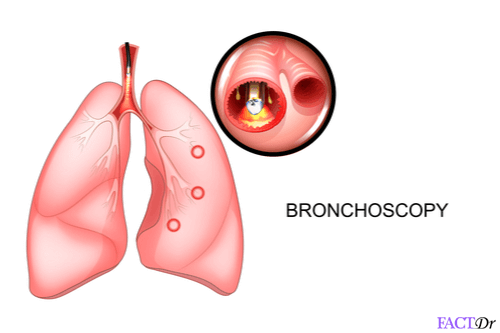
- Bronchoscopy
- Budd-Chiari Syndrome

- Diabetes
- Anatomy of acne: 7 instant steps to kiss those zits goodbye

- Bursitis
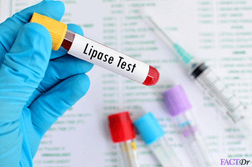
- Lipase Test
- Lactic Acidosis

- Obesity

- Hepatitis
- HB1Ac Test
- Shingles (Herpes Zoster)
- Narcissistic Personality Disorder

- Mad Cow Disease

- Easy and safe tips for ear wax removal you can do at home
- Brainstem Strokes
- Hemophilia

- How to have a healthy pregnancy: Essential tips to remember

- How to correctly perform nasal irrigation using a Neti Pot?
- Porphyria

- Rheumatoid Arthritis

- Low Birth Weight
- Hypertension Profile

- Vitiligo

- Grave’s Disease
- Enlarged Prostate

- Liver Function Test – LFT
- Erythema Multiforme
- Sociopaths: How to identify the ones lurking around you?

- Bromelain: 11 reasons why you should start adding pineapple to your pizza!
- Diabetic Profile

- LDL Cholesterol

- Ducloflex
- Omeprazole

- Missed Period

- Erythritol: A healthy sugar substitute or yet another marketing ploy?

- Autoimmune Hepatitis

- Transverse Myelitis
- Depression

- Pantoprazole
- Bowel Incontinence

- Diplopia

- Pancreatic Cancer

- Agoraphobia

- Varicose Veins

- Undescended Testes
- Angiography
- Autonomic Dysreflexia

- Anal fistula

- Urinary Problems

- 14 go-to foods that are best for a complete natural liver detox

- Anorexia Nervosa

- Pancreatitis

- 12 easy and instant steps to successful weight loss

- Bone fracture
- The ultimate tips on how to boost your brain health

- Measles

- Beginner to Tai Chi? Find the right way to perform this exercise for best results

- Stuttering

- Myopia

- Substance Abuse

- Low carb diet : Go high on fats and low on carbs for a leaner body!

- Peptic Ulcers

- Teething

- Lung Diseases
- Manic depression

- Aspartame – Hidden truths about this storm in a teacup
- Acid Reflux & G.E.R.D.

- Epilepsy

- Dysphagia

- Baldness

- Brucellosis

- Ear pain

- Bloating
- Typhoid

- Kidney Failure
- 18 instant steps to make your jogging routine more powerful

- SIBO Diet: The important dos and don’ts of this gut-healing diet

- Osteogenesis Imperfecta

- Restless Leg Syndrome

- DOMS (Delayed Onset of the Muscle Soreness)

- How to practice yoga for astounding health benefits

- Human Growth Hormone (HGH) Test
- Heart Diseases

- Skin Cancer
- Pregnancy or PMS: The confusion

- Garlic: Nature’s white pill as an effective home remedy
- Cardiac Tamponade
- Ovulation – facts to know about ovulation

- Antibiotic Resistance: How common diseases become deadly
- Bed Wetting In Children

- Your complete guide to an improved and disease-free skin
- Thyroid Disorders
- Cordyceps: How the Caterpillar fungus works as a proven cancer-shield
- Osteopenia
- TMJ – Temporomandibular Joint Dysfunction
- Vitamin D Deficiency

- Constipation

- 5 most effective ways you can lose those pregnancy pounds!

- Weight gain

- What is Qigong and how can it help you channelize you inner “Qi”?

- Testicular Cancer

- Jet Lag

- Psyllium Husk: More than just an effective natural laxative

- Diverticulitis
- Myasthenia Gravis

- Viral Gastroenteritis
- Niemann-Pick disease
- Giant-Cell Arteritis

- Angina
- Amitriptyline

- Premature Birth

- Ankle Osteoarthritis

- Multiple Myeloma
- Stevia: A healthy sugar substitute or yet another risky additive?

- Rotator Cuff Disorders

- Period blood – what it says about your health
- Asperger’s syndrome

- Trigeminal Neuralgia
- Attention Deficit Hyperactivity Disorder

- Water Chestnuts: The tastiest ways to include this healthy tuber in your diet

- Prolactin Test (PRL)
- Cauda Equina Syndrome

- 10 healthy habits: Have a hearty kick-start to the New Year!

- Fevers

- Vagus nerve: 7 health benefits of stimulating the “gut-brain” connection
- Back Pain
- Chloride Test
- Anhedonia: Does the chronic inability to feel joy affect you too?

- Conception

- Whooping Cough

- Cerebral Aneurysm
- Sinusitis
- Pelvic Pain
- Crohn’s Disease

- Thrombophilia
- Pumpkin Seeds: Why pumpkins are more than just for pies!

- Alcoholism

- Ectopic Pregnancy

- Pregnancy

- Type 2 Diabetes

- Avian Flu

- Arthritis

- Autism

- Creatine: Is this the right peak athletic-performance supplement for you?
- Krill Oil: Does the solution to human illnesses really lie at the depths of the ocean?

- Ankylosing spondylitis

- A few facts about Carcinoid syndrome
- Brain Diseases

- Inversion Therapy: The anti-gravity way of healing chronic back pain

- TSH Test – Thyroid Stimulating Hormone

- Myelodysplastic Syndromes
- Bone Marrow Biopsy

- Eczema

- Arrhythmia
- Memory Loss

- Lymphadenopathy

- Acute Pericarditis

- Cardiac Arrest

- Find all the instant ways to get rid of blackheads

- L-Carnitine: The best workout buddy and weight loss supplement you could find
- Gas
- Top reasons why you need to pay attention to your vitamin B6 intake

- Top natural diuretics: The instant benefits you can get from nature’s water pills

- Dehydration

- Botulism

- Rosacea
- Colorectal Cancer
- Edamame: How healthy are these green baby beans?

- 10 reasons why you might be waking up tired

- Vaginoplasty

- Tachycardia

- Dopamine: How dope is the chemistry that governs human emotions?

- Dry cough

- Dandelion: Little parachutes of health and wellness for your whole body

- Epidermodysplasia Verruciformis

- Burns
- Vitamin Profile
- Dyspraxia

- DHEA: Now get unbelievable age defiance with this wonder hormone!

- Gallbladder surgery :Tips on post-operative care and diet

- Seasonal Allergies

- Facts about the Shepherd’s diet

- Kawasaki Disease

- Addiction explained: An obsession that could cost you your life

- Heel spurs
- Liver Cirrhosis
- Fibroids

- Mitral Valve Prolapse

- Amnesia

- Hypothyroidism

- Paleo vs. Keto: Which diet plan is best suited for your body goals?

- AIDS

- Throat Cancer

- Why does the human body experience burnout and how to manage it?

- Asthma

- Dyslexia

- Glucose-6-Phosphate Dehydrogenase (G6PD) test
- Mosquito Bites
- Cardiac Tamponade
- Tremor

- Bipolar disorder

- H. pylori Infections

- Whiplash

- Ovarian Cysts
- Fluoride: An indispensible mineral or a potential toxin ?

- Pap Test: An accurate test that can predict the onset of cervical cancer

- Plexus Slim: Is this pink slimming drink the ultimate one-stop fat burner?

- Melanoma
- Ataxia

- Facial Hair

- Gabapentin

- Actinic Keratosis
- Antiphospholipid (APL) antibody-IgG
- Amino acids: Building blocks of life and a healthy body

- Exercises or diet? Find out the healthiest way to gain weight

- Hiccups

- Anti-Chlamydia Antibody -IgG Test
- Childhood Leukemia

- Mononucleosis
- Herniated Disc
- Bladder training

- Colloidal silver: An essential dietary supplement or yet another pharmaceutical hoax?

- Anaphylaxis

- The complete “what to eat” guide for gluten intolerance
- Tinnitus

- Urinary Tract Infections

- Lauric Acid: 8 health benefits you can derive from coconuts

- Heartburn: What are the exact symptoms and the best home remedies?

- Gingivitis

- The best and safest ways to treat rosacea

- Thyroid Nodules
- Short Breath

- Croup

- Heavy Periods
- Keep your shoulders pain-free with these shoulder exercises
- Cholesterol Management
- Ezekiel Bread: How to make the “world’s healthiest bread” in your own kitchen

- Obsessive-compulsive personality disorder

- Ankle sprain

- Scoliosis

- Antisperm Antibody Test
- Thalassemia

- Coagulation Tests

- C-Peptide test
- Vomiting

- Fructosamine Test
- Don’t choose pain: 10 truths about your back ache (and how to get relief)
- Cancer
- Vertigo

- Frostbite

- Head-injury headaches

- T4 (Thyroxine) Hormone Test
- Tension headaches
- 25-Hydroxy Vitamin D test
- 8 ‘healthy eating’ myths you believed until now

- Malaria
- Cluster headache
- Menstrual headaches

- Prostate Cancer

- Spina Bifida

- Cataracts
- Stop hitting the snooze button: 8 proven steps to perfect sleep

- Asbestosis

- Lazy Eye

- Complement 3 (C3) Test

- Support and care tips for helping people with depression

- Bruxism

- Facts about grass-fed meat

- Acne
- Mono In Teens

- Morning Sickness

- 10 proven ways CBT can help you "untangle" faulty thought patterns
- Carbamazepine / Tegretol test
- Lung Cancer

- 8 reasons why you should grab a bottle of water now!

- Lower Back Pain
- Miracle extract from the root of maca: 10 ways it can help you!
- Cystic Fibrosis

- Cavities
- Valproic Acid Test

- Colic

- Guaranteed ways to lose weight fast and shed those extra pounds!

- Gout
- Neuropathy
- Oxidative Stress: Understanding the most lethal phenomenon happening in your body right now

- Juvenile Macular Degeneration

- Ashwagandha: The best-kept secret of Ayurveda now revealed!

- Menstruation

- Bacterial Vaginosis

- Heart failure
- Exertional headache

- Agave nectar: The bitter truth about this sweetener revealed

- The right way to prepare Alfalfa sprouts at home
- Gatorade: Top things you need to know about this staple energy drink

- Bradycardia
- Sunburn

- Aneurysm

- Hair Loss
- Bunions

- Cilantro: 11 health reasons that will make you wanna use this garnish more!

- Dizziness
- Hyperemesis Gravidarum

- Ulcerative Colitis
- Heel Pain

- Huntington Disease
- 10 Common signs you need to see a mental health counselor now

- Borderline Personality Disorder

- Inhale oxygen, exhale worries: The top breathing exercises and how to perform them

- Pulmonary Embolism
- Transient Ischemic Attack

- Stomach Flu
- Circumcision

- Deficiency & Developmental Disorders: A Complete Guide to Best Remedies
- Breast Cancer

- Stroke
- Hepatitis C

- Progesterone test
- Acute Lymphoblastic Leukemia
- Subarachnoid Hemorrhage
- Polio

- Your Complete Guide to Preventing Birth Defects

- COPD-Chronic Obstructive Pulmonary Disease

- Plague
- Muscle relaxers: Think twice before popping prescription drugs for muscle pain

- Encephalitis

- Avascular Necrosis
- Toxic Shock Syndrome

- 6 arguments in favour of glucosamine and where can you find it
- Miscarriage
- Sleep Disorders

- How cooking with CLA safflower oil can help you lose weight?

- Beware of these common foods that cause high bodily inflammation!

- TMJ – Temporomandibular Joint Disorders

- Spinal Fracture
- Hibiscus: Nourish your beauty and health with these vibrant blooms

- Bone marrow aspiration

- Binge Eating Disorder

- Septic Arthritis
- Sex Hormone Binding Globulin (SHBG) Test

- Unbelievable health benefits of gooseberries (and a few tasty recipes)

- Metatarsalgia

- Black Seed Oil: What secret benefits could it bestow on your health?
- Valerian Root: The best way to achieve tranquil sleep every night

- Brain Cancer

- Splenda
- Sushi: health benefits and recipes

- Dental Care

- Vaccines

- Cerebral palsy

- Sciatica pain relief: Get rid of the pain with these easy daily tips
- Halotherapy: How inhaling salt can work wonders for your body
- Beware: Ignoring these tell-tale signs of stroke could cost a life
- Hay Fever

- Pilonidal sinus

- Bulletproof Coffee – Breakfast of champions or yet another health fad?

- Antiphospholipid Antibody(APL)-IgM test

- Gastroenteritis

- PiYo (Pilates + Yoga): Bringing you the best of both the worlds

- Gout Diet : Get relief from painful joints by eating these foods

- Allergies

- Schizophrenia
- Leukaemia

- Cold and Flu

- Contraception

- Manuka Honey: What is it and why should you choose this over regular honey?

- Anxiety and Panic

- Best treatment for piles in Bangalore
- Kale: With more iron than beef, this superfood is the ultimate way to lose weight

- Anti-Social Personality Disorder

- Apple cider vinegar: Weight loss, shinier hair, and more!

- Hot cups for detox? Read all about the ancient technique of cupping

- Arthritis Profile
- Legumes: Little pods of infinite nutrition you should consume daily

- Spirulina: Battle diabetes and cholesterol with this blue-green algae
- Night Terrors

- Leprosy

- Knee Pain

- Menopause
- Winter skin hazards
- Tennis Elbow

- Iron-Rich Foods: Eat these foods to keep fatigue and anemia away!

- SARS
- Sore Throat
- Blindness

- Air Fryer: The ultimate savior of fried food addicts!
- Metoprolol

- Ventricular diseases
- Himalayan salt lamps: Do you want to bask in the pink glow of health?

- Troubled with ear infections? Find easy and instant home remedies here
- Seasonal Affective Disorder

- Hemp Seeds: The actual science behind this controversial crop

- Heat Stroke
- Avocado: 15 reasons why this fruit should be a staple in your diet

- Glutamine: Why is this the right choice for muscle-gain supplement?
- Keratosis Pilaris

- Insomnia

- Green, brown, & bitter: The instant advantages of including fenugreek in your diet
- Sensory diabetic neuropathy
- Chronic Fatigue Syndrome
- Perimenopause

- Coughs

- Pelvic Ultrasound

- Quinoa : A prodigious superfood you need to include in your diet today!

- Water Fasting: What are the pros and cons of this practice?

- Fibromyalgia

- Panic Attack

- Stress

- Anti-Hepatitis B Core Antigen (AHBc) IgM
- How to beat the common cold, flu, and sinus infection instantly?
- Narcolepsy

- Premenstrual Syndrome (PMS)

- Beta-alanine: A powerhouse of proteins to charge up your workout

- Chest Pain
- Biotin: One-stop-shop for shiny hair and an active body!

- Aortic Dissection
- EMG- Electromyography

- Summer Skin Hazards
- Chronic Lymphocytic Leukemia
- Coronary Artery Disease

- Drug Overdose

- Shoulder Fracture

- Vital Organs & their diseases: A Complete Guide to Best Treatment and Remedies

- Swine flu

- Preeclampsia

- Dates: The complete list of instant health benefits this dry fruit can bestow

- Deflazacort
- Chikungunya-IgM Test

- Snoring

- We bet you didn’t know that turmeric could be this beneficial!

- Ginseng: Top reasons how this root can help you boost energy ( & libido too!)

- Latest findings on Kombucha; a teacup full of yeast and bacteria

- Cardiolipin Antibody (ACL) –IgG Test

- Migraine

- Is it possible to achieve full body detox with Master Cleanse?

- Hypoglycemia

- A sneak peek into the top 5 deadliest cancers and the true nature of their origins

- Tourette's Syndrome
- Diabetic foot ulcer
- ACL Injury

- MCT Oil : The kind of fats you should be eating

- Triple Marker Test

- Aging
- Castor oil : A vitamin-rich oil for shiny locks and healthy bowels
- Sleep Apnea

- Hot Flashes

- Fatigue

- Shilajit: A miracle Himalayan herb for an active and happy life

- Bad Breath

- Acute Myeloid Leukemia

- Carpal Tunnel Syndrome
- Ménière’s Disease
- Rett syndrome

- Fanconi anemia
- Parkinson’s Disease
- Chikungunya Fever

- Atorvastatin
- Sciatica

- Triglycerides: The most effective steps in bringing the levels down

- Spondylitis

- What is the exact job of an OB-GYN and why should you consult one?

- Spider veins

- Caffeine-related headaches

- Bladder Cancer
- Headache

- Psoriatic Arthritis
- Echocardiography – ECHO
- Inflammatory Bowel Disease

- Don’t let your hearing dip with age. Here’s what you need to do

- Listeria

- Low progesterone: The dangers of this hormonal imbalance and how to correct it

- ALP test – Alkaline Phosphate
- Ovarian Cancer
- Is this is the real life? Is this just fantasy? – Decoding the myths of lucid dreaming

- Corn: Be amaized by the health benefits of including it in your diet

- Glaucoma

- Male pattern baldness

- Amyotrophic Lateral Sclerosis (ALS)

- Brain Arteriovenous Malformation

- Alzheimer’s

- Prediabetes

- Gua sha: Scraping away layers of pain with this ancient healing tool

- Overactive Bladder
- DASH Diet: What to eat in a “Dietary Approach to Stop Hypertension”?

- Testosterone: And you thought it was just brawns and sex drive

- Seborrhoeic Dermatitis

- Infertility profile (women)

- Endometriosis
- Gestational Diabetes
- Hearing Loss
- Amaranth: Why is this native Peruvian grain called the “crop of the future”?
- Multiple Sclerosis
- Creatinine Test

- Insulin Resistance

- Parkinson’s stages

- Hypertension
- Congestive Heart Failure

- Zika Virus Infection

- Achilles Tendon Injuries
- 5-htp: Is this the ultimate cure for those pesky migraines attacks?

- Amnesia: It’s not just a blow on the head that can erase your memory

- Ginger: Why is it called the root of all remedies?

- Psoriasis
- 10 reasons why you should up your selenium intake
- Deadly infections guide: Find the best & safest cures for them here

- Common mental health disorders :The silent stigma surrounding millions

- Sore Throat: How to treat this throat infection at home?

- Your Complete Guide to Oral health: Best supplements for your teeth & gums

- Vitamin B12: The unknown repercussions of this nutrient deficiency

- Post Traumatic Stress Disorder (PTSD)

- Cardiolipin Antibody (ACL) – IgA test

- Tomato: All you need to know about this juicy vegetable (or a berry?)

- Nipah Virus Infection

- Amyloidosis
- Tendinitis
- Pituitary Gland Disorders
- Anti-Inflammatory foods: 18 foods to a disease-free you

- Greek Yogurt: Discover the secret ingredient to the real Spartan strength

- Neck Pain

- Prostate-Specific Antigen (PSA) Test

- Burpees: World’s most challenging fitness routine now made simple for you

- Attention Deficit Hyperactivity Disorder in Children

- Anthrax

- Lead Poisoning

- The dangers of having insufficient digestive enzymes (and how to pump these naturally)

- Probiotics: How to best feed your gut bacteria for higher immunity

- Hydrochlorothiazide
- Prednisone

- The tell-tale signs of pregnancy you should know

- Top foods with polyphenols and why you should eat them

- Foot Problems

- Vitamin D foods: Know what to eat for stronger bones and a cancer-free body
- Medication-Overuse Headache
- Gingko Biloba: Therapeutic lessons from the world’s oldest tree

- Bronchiectasis
- Azithromycin
- Lewy Body Dementia

- Tea Tree Oil : The one stop cure for skin ailments and more…

- 15 Signs on your face you should never ignore

- Histoplasmosis
- Propolis: Yet another amazing gift by the bees to the humans

- What are the common mental health disorders in children and teens?
- Coconut Oil: How one oil dominates the kitchen and the medicine kit too

- Microneedling: Does this cosmetic procedure live up to the hype?
- Chronic Pelvic Pain

- ADD versus ADHD: What are the fundamental differences?

- Carotid Artery Disease

- Psychosis

- Sharpen your brain with these top brain-friendly foods!

- Irritable Bowel Syndrome

- Infertility

- Celiac Disease

- Newborn Screening Tests

- Urinary Incontinence In Women

- Glycemic Index

- Head Lice

- Easy and effective ways to get rid of the stubborn belly fat

- Sensory Processing Disorder
- Female Pattern Baldness

- BCAA: Now get more out of your workouts with this miracle protein

- Hemoglobin Variant Analysis
- Beets: Top reasons why these nutrient-dense roots should be a staple
- Sinus Headaches
- Geriatric health: Do you still think age is just a number?
- Pain Management
- Whole30 Diet – Why eliminating certain foods just doesn’t cut it

- Aphasia

- Ondansetron

- Metabolic syndrome

- Cottage cheese: What can you find in this healthy chunk of fresh milk cheese?

- Endoscopy

- Rising mercury and your health: How Global Warming is damaging your body

- Age-Related Macular Degeneration

- Anti Hepatitis-B-Surface Total Test

- Dementia

- Type 1 Diabetes

- Osteoarthritis
- Evening Primrose: 13 incredible ways this omega 6-rich oil can transform your body

- How to safely use leptigen as a miracle weight loss supplement?

- The complete fitness guide on how to perform Tabata

- Neurologist: How can the specialist help you avert fatal diseases

- Serotonin: What you didn’t know about this “feel-good” chemical

- Bone Scan

- Lyme Disease

- 8 appetizing and mouth-watering recipes that every low-carb dieter should try

- 9 reasons why almonds should be consumed daily
- Anti-Hepatitis B Core Antigen (AHBc) Total

- Bone Density Test
- Anti Mullerian Hormone (AMH) Test

- Spinal cord tumors
- Keto Diet: Did you know you could eat fats to burn fats?

- Allopathy vs homeopathy vs ayurveda treatment for piles, fissures, and fistula

- Liver Biopsy
- Sweeteners – Are They Good or Bad for Our Health?
- Chia seeds: How to consume this staple Aztec food to get a leaner body
- Tattoo Aftercare: The best practices to follow once you get inked

- Low carb vegetables: A complete list of what to pick from during a keto diet

- Oil Pulling: The instant benefits of this ancient Ayurvedic cleansing technique
- FSH (Follicle Stimulating Hormone) Test
- Nise
- Concussion
- Canola oil: How safe is it to consume this popular cooking oil?

- Botox: Top things you should know before undergoing the procedure
- Pomegranate: Why are they called ruby red pearls of health?

- Abdominal Ultrasound

- Lithium test

- Kshar sutra treatment for piles, fissures and fistula

- Element 22 test (Nutrients & Toxicity)

- Potassium: How only 100 mg a day can dramatically increase your lifespan!

- Leaky Gut

- Spinal Stenosis

- Herpes simplex virus (HSV)-IgG Test

- Complement 4 (C4) Test
- Proven health benefits and nutrition of macadamia nuts

- Obstructive Sleep Apnea

- 9 amazing ways chondroitin can help you bounce back to health
- Quercetin: The ultimate anti-inflammatory plant compound

- Essential oils: Choose the right one based on your need

- Everything you need to know about COVID-19

- Generalized Anxiety Disorder

- IME9
- Tonsillectomy: When do you actually need to get your tonsil glands removed?
- Nootropics: How safe are these drugs that make you smarter?

- High protein diet: How much protein is too much protein?
- What does hypoglycemia look like when you don’t have diabetes?
- Toxo Gondii-IgM Test

- Anticholinergics: The diverse range of diseases a single drug can easily treat
- Whey Protein: The real reason why this supplement is a must for bodybuilders
- Unconjugated Estriol (E3) test

- Dark, hot, & steamy: The remarkable health benefits of drinking coffee

- Plantar Fasciitis

- A few details about Biltong

- Biopsy

- Rick Simpson’s Oil: The truths and controversies of this cannabis product

- Breast augmentation

- The features of an Optavia diet

- Bone Broth: Why is this simmering bowl of soup called the “Jewish Penicillin”

- 10 fantastic Superfoods you need to include in your diet from today!

- Matcha: The unthinkable health benefits of this unique green tea
- Glutathione: Probably the strongest antioxidant known to mankind

- Olive Leaf Extract: The healthy goodness of olives in a capsule

- THC: The science behind why marijuana is extremely addictive (and toxic) for you

- Elderberry: More than just a folk medicine for cold and sinusitis

- TRX: Why is this total resistance workout the best choice for your body?

- Calorie Counter: Count your calories before they flab

- Graston technique: A faster way to accelerate your rehabilitation after injury

- Why is olive oil probably the healthiest cooking oil you will find

- Aloe Vera: The amazing things you can do with this antioxidant-rich gel
- Infertility profile (men)

- Max thalassemia Profile

- Labiaplasty

- Military Diet : Why following a highly restrictive diet is a bad idea

- Free testosterone
- Experts believe these are the best protein foods for you

- Chicken fajitas – A delicacy to fix weeknight dinner dilemma!

- Diabetes in women: Understanding the dangers, risk factors, & treatment
- Grapeseed Oil: Find out how to use this oil without any side-effects

- Intermittent Fasting : The “real secret” behind healthy weight loss?

- The best pre-workout supplements to help you overcome gym fatigue

- Is shingles contagious? If yes, then how can you prevent this infection?
- Top Natural Repellents for Mosquitoes with negligible side-effects

- Cystoscopy

- Total Cholesterol Test
- Walking Pneumonia

- Global infections scenario : Which are the most common ones?

- Cortisol Test

- Fructose: Healthy fruit-product or loaded with toxic calories?

- Fish Oil: Why do doctors love prescribing these to heart patients?

- Echinacea: Why is this ancient herb re-emerging as an all-rounder supplement?

- The most effective tips on how to stay awake at work

- The best lower back strengthening exercises for freedom from chronic pain

- Cytomegalo Virus (CMV) – IgM Test

- Holistic Healing: How does it harmonize your mind, body, and soul?

- Vitamin C: The instant health benefits of adding the citrusy goodness to your diet

- Homocysteine Test

- How to spot the early signs of lung cancer?

- Atrial Fibrillation

- Paleo snacks

- Beta-Thalassemia Screening

- Clostridium difficile infections
- Guaranteed tips on how to bounce back from low testosterone levels

- Buckwheat: Instant health benefits of adding this super healthy grain in your diet
- Kratom: Why is this distant cousin of coffee banned in numerous countries?

- Apolipoprotein B-(Apo-B)

- Pregnancy – Early signs and symptoms

- 18 amazing uses of Baking Soda you didn’t know!

- Positron Emission Tomography – PET Scan

- A few facts about threatened abortion

- Beta-carotene: Top reasons why you need to include vitamin A in your diet

- Naturopathy: When the goodness of nature heals you

- Collagen: Get age-defying skin and faster hair growth with this miracle protein

- Calories: How to keep a check on these for faster weight loss

- Postpartum Depression

- Influenza A

- Nitric oxide: Why is this heart-healthy supplement every sportsman’s first choice?

- Kidney stones

- How chlorophyll is more than just a green pigment meant for photosynthesis

- Treponema Pallidum Hemagglutination Test

- Hypertension: The best ways to prevent the onslaught of this mass-killer
- Magnesium Test

- Luteinizing Hormone (LH) Test

- Blood in your stool

- Lecithin: Learn how you can utilize this fatty acid to improve your cholesterol count

- Amniocentesis

- Medical Mythbusters : How many of these myths did you think were true?

- Top reasons why broccoli is a high-ranking powerhouse vegetable

- Active Release Technique: Say goodbye to chronic body pain with ART

- Ketosis: A potentially dangerous side effect of the popular keto diet?

- Green Tea: Does it really aid weight loss and reverse aging?

- Goldenseal: Experts reveal why it is the most useful herb known to mankind
- The hidden dangers of magnesium deficiency and how you can prevent it
- Citrate or Oxide: Which magnesium supplement is best for your body?

- Acupuncture: 7 remarkable ways these pain-free needles can bring you holistic healing
- Omega 3: Now you know why fats are healthy for you
- Experts reveal, how long does marijuana actually stay in your system…

- If it fits your macros (IIFYM) : A contemporary macro diet regime

- MRI – Magnetic Resonance Imaging

- Benefits of exercise: Top reasons why you should be sweating it out more in the gym

- Iodine Deficiency: Risks and complications and how to avoid them
- Peanut Butter: A spread of nutrients on your morning toast
- HCG Diet: The dangers of using this diet to lose weight

- Alpha Feto Protein (AFP) Test

- Termination of pregnancy

- Migraine: Get rid of this agonizing pain naturally with these home remedies
- Rhubarb: The secret nutrients hidden in these deep red stems

- Cranberry: Top reasons why you should grow this “superfruit” in your own backyard

- Tanning booths: To tan or not to tan, that is the question
- Sigmoidoscopy

- Night sweats

- Cannabidiol benefits: Evaluating the “highs” of CBD Oil

- Proteins: What is the right protein-intake for your specific needs?

- Paleo profile
- Difference between piles, fissures, and fistula
- Pregnancy test – Urine & Blood

- Best Weight Loss Pills: How to choose your drug wisely
- Activated charcoal: The instant health benefits of this “diamond in the coal mine”
- Organic Foods: Is the popularity of pesticide-free food justified?

- Yerba Mate: Could the national drink of Argentina successfully replace coffee?

- Did you know vitamin E can fight these fatal diseases?
- Are you sure you are drinking enough water?

- Hyaluronic Acid: The real helper chemical for younger skin and stronger bones

- Mammography

- Electric toothbrush vs manual toothbrush: Who wins the dental war?

- Iron Deficiency: 7 major side-effects and steps to overcome them

- From treating insomnia to joint aches, discover how eucalyptus extract can help you

- Melatonin: Fascinating facts on the hormone that brings you the Zzzs

- Colonoscopy
- Cinnamon: Sprinkle this on your morning coffee to gain guaranteed health benefits

- The top natural antihistamines and how to use them

- Best ideas for pregnancy snacks

- Forskolin: Did you know you could use this mint-like extract for weight loss?

- Spinach: The ultimate way to boost health with this green superfood!

- All that you should know about Roseroot

- Comprehensive Metabolic Panel
- Steroid Profile

- High blood pressure during pregnancy: Causes and remedies

- Lavender: The calm and soothing oil your body needs right now

- Prebiotics: Eat the foods your gut bacteria love eating

- Anti-Chlamydia Antibody IgM

- Laughter Therapy– The Best Medicine for Healthy Living

- L-arginine: Your search for the ultimate fitness booster ends here

- Vitamin D3: Speed up your journey to a fit body and mind

- How to make the apple cider vinegar diet work for you?
- Dr. Gundry’s diet: A new concept in dieting

- Pilonidal sinus

- 12 instant and most effective remedies for a hangover

- Best keto snacks to munch on when you are on a keto diet

- Top 15 fat burning foods you need to start eating from today!
- Garcinia Cambogia: Combat weight gain and obesity with this effective natural supplement

- High global burden of common widespread health conditions

- How does Reiki actually work and what are its benefits?

- Occupational Exposure & Lymphomas: A Complete Guide to Dangers & Remedies

- Testosterone test

- Estradiol Test

- 11 natural ways to cure upper respiratory tract infections

- The Vegan Diet: The benevolent way of eating that wins hearts (and tummies)

- Hydrocele
- Golden Milk – Your Recipe to Good Health

- Best Multivitamins for men: Find the ones best suited for you

- MSM: An effective sulfur supplement to ease those aching joints

- Why calendula extract ought to be part of your personal health kit?

- Hate making breakfast?Fast and healthy breakfast recipes just for you!

- Transcend deeper into a healthier mind and body with transcendental meditation

- Children Nutrition and Toxicity Profile
- Can’t eat eggs? Here are the best egg substitutes for you

- Practical Motherhood Tips for Working Women

- Do you have COVID-19? Know the critical signs and symptoms
- T3 Test
- Aromatherapy: Can this sense-stimulating therapy help you?

- Gluten-free diet – a comprehensive, easy to follow guide

- Doppler Ultrasound

- Septoplasty

- ERCP – Endoscopic Retrograde Cholangiopancreatography

- Adenosine Deaminase Test

- 12 myths and facts about anal piles

- Ultrasound

- Rhodiola Rosea: Hoist yourself to high stamina and a stress-free brain

- PCOD(Poly Cystic Ovarian Disorder) Profile
- Root canal

- Folate: What are the best ways to add this nutrient in your staple diet?

- Diabetic Retinopathy

- Experts believe these are the healthiest diets for you
- 10 science-backed reasons why salmon is probably the healthiest fish known

- Best Practices to Prevent and Control COVID-19

- Paleo Diet: Is eating like the Caveman the right way to go?

- What should you know before anal fissure surgery?

- How the deficiency of Coenzyme Q10 could bring your organs to a standstill

- What can you do if you are COVID-19 positive: Practical coping tips

- HPV Digene Hybrid DNA Detection test
- Calcium test

- Meditation: The shortest route to complete peace of mind (& body) in today’s world

- Is there more to your chronic fatigue than just tiredness?

- Toxo Gondii-IgG Test

- Inner Knee Pain

- Tips to take care of your mental health during COVID-19

- Is Chiropractic really effective for relieving back pain?

- Buddha bowl

- Immunoglobulin A Test

- Anorectal disorders – laser surgery or home remedies?

- COVID-19: Know all about types of diagnostic testing, procedure, risks

- What is Baby Led Weaning and how does it compare to traditional weaning?

- Does your teeth really need charcoal toothpaste?

- Revision Total Knee Replacement surgery

- Chemo brain

- Burn those years of stubborn fat-deposits with a few minutes of HIIT!

- Egg freezing: Questions every woman has before preserving her eggs

- What makes Homoeopathy a safe way towards recovery?

- Ceruloplasmin Test

- Immunoglobulin G Test

- Lipoma
- How excess sugar changes your body over time

- Diabetes Diet 101: All the foods you should eat ( & avoid)
- Diabetic foot ulcer
- COVID-19: What is the treatment and how long is the recovery?
- COVID-19: Possible serious long-term side-effects for patients

- Hormone Replacement Therapy-Symptoms, Benefits, Types, Side Effects, Lifestyle Changes

- Medical conditions yoga can help with

- Is fecal transplant a safe method of gut disease treatment?

- Varicose veins

- Rhinoplasty
- Breast Reduction Surgery

- Ear Surgery

- Breast Augmentation

- Safety tips and precautions during COVID-19

About
Trending Topics
Top Stories
- Blue Balls
- Ferritin Test
- Rubella IgG Test
- Tonsil Stones
- Enterogermina
- Muteness
- Random Blood Sugar Test
- Sebaceous Cyst
- Lectin: The common link of proteins between peas and people
- Augmentin 625
- Lipid Profile
- Primolut N
- Bifilac
- Cervical Cysts
- Anti-Thyroglobulin Antibody test
- Pulmonary Function Tests
- Lupus Rash
- Pott’s disease
- Dexorange
- FBS Test – Fasting Blood Sugar
- EKG – Electrocardiogram
- Rheumatoid Factor (RF)
- Flunarizine
- Tongue infections
- Leukoplakia
- The real reason why you shouldn’t be eating ramen noodles
- Skin Rashes
- Betadine
- TBHQ: A carcinogen lingering in your child’s favorite snacks
- Post-Prandial Blood Sugar
- T Bact Ointment
- Thyroglobulin (TG) Test
- Dyshidrotic eczema
- Serum Electrolyte
- Becosules
- Passion fruit: How one exotic fruit can help you fight infections and cancer!
- Hydrocele
- Beriberi
- Orofer XT
- Chondromalacia
- Thyromegaly
- Cervical Polyps
- Amoebiasis
- Blue Waffle Disease
- High platelet count
- Hepatitis B Envelope Antigen (HBeAg)
- Avil
- Treponema Pallidum Antibody(TPAB) test
- Arterial Blood Gas Analysis
- Nurokind LC
- Inverted nipples
- AST- Aspartate Aminotransferase Test
- Serum Zinc Test
- Herpes Simplex Virus I (HSV)-IgG Test
- Chiggers
- The importance of roughage in diet
- Evion LC
- Hangnails
- Duphalac
- Cherry Angioma
- Diverticulitis diet: The right way to eat if you suffer from the disease
- Wasp Sting
- HLA-B27 test
- Myospaz
- Balanitis
- Non-Hodgkin’s Lymphoma
- Free Triiodothyronine (FT3) Test
- Abscess
- DHEA Sulfate (DHEAS) Test
- Chromium Toxicity
- Hypermetropia
- Betnovate
- IgE test – Immunoglobulin E
- Pus
- Abdominal CT scan
- Pellagra
- Metabolic Disorders
- Yellow poop
- Autoimmune Diseases: Find out if your body is attacking you right now
- Cardiolipin Antibody (ACL) –IgM Test
- Dengue NS1 test
- Beta 2 Glycoprotein 1 IgG
- Vitamin K2: 8 reasons why you need this bone-building & cancer-fighting nutrient
- Fibroadenoma
- Prickly Heat Rash
- Silicon Dioxide: How can a component of sand be essential to your wellbeing?
- Maltitol: Things you must know about this artificial sweetener
- Albinism
- Enlarged Liver
- Racecadotril
- Viral Infections
- Zerodol
- Urinary Microalbumin Test
- Helicobacter Pylori – IgG Test
- Erythropoietin (EPO) Test
- Endomysial Antibodies (EMA) Test
- Fatty Liver Disease
- Brown Recluse Spider Bites
- Taxim O
- Shelcal 500
- Ketorol DT
- Kidney Cysts
- The Big 5 lifestyle diseases: How your everyday living might be killing you
- Excretory System Diseases
- Smallpox
- Gum Disease
- Clavam 625
- Pan D
- Swollen Lymph Nodes
- Bilirubin Test
- Sickle cell disease
- Klinefelter’s Syndrome
- How do neutrophils protect you from fatal bacterial attacks?
- Chymoral Forte
- Brain Cysts
- Cavernous Sinus Thrombosis
- Intestinal Adhesions
- Smegma
- CLA: A breakthrough weight loss supplement with minimal side effects
- Birthmarks
- Cytomegalovirus (CMV)- IgG Test
- Eustachian Tube : Functions and top home remedies to prevent its infection
- Free thyroxine test (FT4)
- Chromium Picolinate: An essential mineral supplement for faster weight loss
- Alexandria’s Genesis
- Skin Tags
- Cellulitis
- Antinuclear Antibody Test – (ANA)
- Enteritis
- Genital Herpes
- Maltodextrin: What are the hidden health benefits of this food additive?
- Ingrown Toenail
- Dry skin
- Cheilitis
- Phlebitis
- Poop chart: Top things you didn’t you your poop could reveal about you
- Occupational Hazards: How to vouch for your health at your workplace
- Mox 500
- Levosulpiride
- Anti-CCP Test – Anti-Cyclic Citrullinated Peptide
- Disodium Hydrogen Citrate
- Genital Warts (HPV)
- Myocardial infarction
- Benign Tumours
- Scabies
- Jock Itch
- Hydrocephalus
- Zerodol SP
- Prolapsed Uterus
- Duphaston
- BRAT Diet: What is the right way to follow this diarrhea-relieving diet?
- Tissue Transglutaminase Antibody (tTG) Test
- Internal Bleeding
- Hepatitis Profile
- Etizolam
- Deep Vein Thrombosis
- Burning mouth syndrome
- Zifi 200
- Anti Ds-DNA antibody Test
- Norflox TZ
- Abnormal Vaginal Bleeding
- Cystoscopy
- Neural tube defects
- Salivary Gland Infection
- Electroencephalogram – EEG
- Sputum test
- Rectal Prolapse
- Gonorrhoea
- Cardiac Profile
- Top 6 remedies to treat a razor burn at home
- Temper Tantrum
- Foot Corns
- Asthenia
- 17 OH Progesterone test
- Nexito Plus
- Pinworms
- Poison oak
- Pyelonephritis
- Stye
- Lisp
- Bronchitis
- Dysentery
- Mumps
- Moon Facies ( Cushing Syndrome)
- Eating Disorders: Lifestyle choice or a psychological condition?
- Scalp Psoriasis
- Dwarfism
- Typhus
- Folate test
- Hypersplenism
- Rubella
- Hiatal Hernia
- Skin irritation
- Endocrine System Disorders
- Gilbert’s Syndrome
- Razo D
- Ringworm
- Athlete’s Foot
- Blood blister
- Deafness
- Fungal infections
- Hives
- Swollen Feet
- Helicobacter Pylori – IgA Test
- Genetic diseases
- Hemolytic Anemia
- Tonsillitis
- Arachnoid Cysts
- Carrageenan: How a simple seaweed extract could better your gut health & immunity
- Amylase Test
- Cerebral Cavernoma
- Flagyl 400
- Anti-Microsomal Antibody AMA Test
- Immunoglobulin M Test
- Common diseases that could cripple your vital organs
- Emphysema
- Anencephaly
- Proctitis
- Androstenedione Test
- Drotin DS
- Oxycodone
- CBC (Hemogram 6-part diff) blood test
- Oral Glucose Tolerance Test – GTT
- Stye : The best natural home remedies to ease the pain
- Piaget stages: Do they accurately describe the way the human brain develops?
- Lipoma
- Heat Rash
- Costochondritis
- Trisodium Phosphate: How a paint thinner made its way into your breakfast
- The science behind daith piercing: Can it really cure migraine?
- Copper Serum Test
- Arm fracture
- West Nile Disease
- Leukocytosis
- Maladaptive Daydreaming
- Tonometry
- Troubled with IBS? Here’s a complete roadmap to the low FODMAP diet
- Ulcers
- Panera Bread: The truth behind this “healthy” restaurant chain
- Ciplox Tz
- MRSA – Methicillin-Resistant Staphylococcus Aureus
- Beta hCG Test
- Sepsis (Blood Poisoning)
- Cloudy Urine
- Hematuria
- Free PSA Test
- Blood Disorders
- 9 benefits of walking we bet you didn’t know!
- Stuffy Nose
- Sitophobia
- Squid Ink: A unique food coloring and flavoring agent
- Oral Leukoplakia
- 5 unbelievable effects of dance on your overall health!
- Hand Fracture
- Werner’s Syndrome
- Dislocated Jaw
- Acanthosis nigricans
- Syphilis
- How are BMI and BMR different and what do these numbers mean?
- Blood thinners
- Rickets
- Adenoiditis
- Sinarest
- Ascites
- 8 hidden causes of obesity you probably didn’t know!
- Giardiasis
- How can berberine supplements help you live longer and healthier?
- Freckles
- Spider Bites
- Comedones
- Enlarged Heart
- Albumin Test
- Varicocele
- Milk Thistle: Find possible cures for fatal diseases in these purple blooms
- Tongue cancer
- Caralluma Fimbriata: How to eat this cactus to lose weight
- Anemia Profile
- Trichomoniasis
- These are the top foods to increase your hemoglobin count
- GGT – Gamma-Glutamyl Transferase Test
- Hookworms
- Quadriplegia
- Wound debridement and dressing
- Don’t let your sleep deficit grow into memory loss or heart attack
- Oligohydramnios
- Pre-cancerous Skin Lesions
- Lipoprotein (A) Test
- Lockjaw
- CA 15-3 Test
- Baby Acne
- Atelectasis
- How accurate is the hair follicle drug test?
- Congenital Glaucoma
- Canker Sores (Apthous Stomatitis)
- Beta 2 Glycoprotein 1 IgM
- Rhabdomyolysis
- Yellow Jacket Sting
- The real reasons for your mood swings and how to overcome them
- Charley Horse
- Power up your gut: 8 proven steps on how to improve your digestion
- Microcephaly
- Blisters
- Are you depressed or just stressed? Know when to see a doctor
- Addison’s Disease
- Anal Abscess
- Motion Sickness
- Folliculitis
- Dyscalculia
- LP-PLA2 Test
- Tongue Diseases
- Ketoacidosis
- Plantar Warts
- Ingrown Hair
- Anhidrosis
- Edema
- Meftal Spas
- Sustan 200
- Chlorella: Therapeutic powers of the billion-year-old algae now within your reach
- Lipodystrophy
- Thalassemia
- Xylitol : Is this the right sugar substitute for you?
- Brain-Eating Amoeba (Naegleriasis)
- Signoflam
- Oxalates: How healthy greens can sometimes be bad news for your kidneys
- Staph Infection
- Green Healing: The amazing health benefits of being in nature
- Bruises
- What are Shiitake Mushrooms and why should you eat them?
- Cheston Cold
- Skin Problems
- Jicama: A fiber-rich tuber for your weight loss goals
- Bone tumor
- Silicosis
- Is bronchitis contagious? And how to prevent this infection?
- Insect Bites
- Chicken Pox
- Otitis media
- Blood Element Analysis Test
- Jackfruit: Slice your way into higher immunity, better digestion, & more…
- Joint Damage
- Carcinoembryonic antigen (CEA) test
- Appendicitis
- 7 liver detox facts and why ignoring these could kill you!
- Jaundice
- Green poop : What are the reasons and how can you correct it
- Hyperthyroidism
- Herpes Simplex Virus (HSV)-IgM Test
- Hypocalcemia
- Vitamin B12 Test
- Skull Fracture
- Astaxanthin: Why experts believe it could be 550x more potent than vitamin E!
- Itching
- Poison sumac
- Thyroid Scan
- Quinine: How gin and tonic came to be known as a potent health elixir
- Flat Feet
- Dengue Fever
- Itraconazole
- Pneumothorax
- Syncope
- Connective Tissue Diseases
- Colloid Cyst
- Molluscum contagiosum
- Skin Problems in Children
- Contact dermatitis
- Pneumonia
- Speech disorders
- Night Blindness
- Non-Allergic Rhinitis
- Lupus
- Subdural Hematoma
- Coconut sugar: What is it and is it healthier than table sugar?
- Nail Fungus
- CT – Computed Tomography Scan
- Iron Deficiency Profile
- Dragon Fruit – Blessing from Nature’s Basket
- Stages of wound healing
- Bulimia Nervosa
- Cold Sores
- Combiflam
- Quadriceps: Why stretching these is a must before going for a run
- Endometritis
- What to eat, what not to eat in the South Beach Diet?
- Bacterial Pneumonia
- Ear Infections
- 5 things you didn’t know about balneotherapy and how you can do it at home
- Scurvy
- Xanthan Gum: The pros and cons of this synthetic gummy additive
- Colon Polyps
- Tuberculosis
- Anemia
- Metal toxicity
- Arsenic Poisoning
- Surgery or home-remedy; what is the right way to treat a burn
- Hodgkin’s Disease
- A complete list of low-fat diets and how to follow them
- Lymphoma
- Trypophobia
- Hysteroscopy
- Fibrocystic Breast Changes
- Juice Plus: Can you really replace actual fruits with pills?
- Cystatin C Test
- Down syndrome
- Gastritis
- Stretch Marks
- Peripheral Artery Disease
- Pernicious anemia
- Pain disorder
- Yeast Infection
- Pressure ulcers
- Bell’s palsy
- Hymenoplasty
- The actual science behind smelling salts and how it helps in injury recovery
- Burkitt Lymphoma
- Cat-Scratch Disease
- Rheumatic Fever
- Bubble Tea: Bubbling with nutrition or a hidden sugar mine?
- Portal Hypertension
- Hyperventilation
- Temporal Arteritis
- L-Theanine: Better sleep, higher focus, and many other benefits
- Kava: Is it the healthy substitute for alcohol you have been waiting for?
- Postpartum Infections
- Traumatic fractures
- Tamarind: The top health benefits of this staple Asian ingredient
- Keratitis
- Chest X-Ray
- Chlamydia
- Thrombocytopenia
- Dysgraphia
- Sugar: 7 new shocking truths revealed about the sweet poison
- Immune System Disorders
- Sodium chloride: The wonders and dangers of the simple table salt
- Rabies
- Nutritional Yeast : How can a fungi culture be good for your body?
- C-Reactive Protein Test
- Muscular dystrophy
- Pelvic Inflammatory Disease
- Empyema
- Eye-sight problems
- Viral Meningitis
- Liver Cancer
- Pseudocyst
- Esava
- Itchy Scalp
- Enlarged adenoids
- Conjunctivitis
- Neonatal Jaundice
- Hydroquinone: Is it the best topical treatment for hyperpigmentation?
- Painful Urination
- Arteriosclerosis
- Bloom Syndrome
- Herpes Simplex Virus I (HSV)-IgG Test
- Cervicitis
- Appendix pain: Recent findings on this not-so vestigial organ
- Cyclopam
- Poison Ivy
- Myelography
- Nursemaid’s Elbow
- 11 unbelievable health benefits of thyme you didn’t know!
- Experts reveal the safest ways to perform an enema at home
- Osteomyelitis
- Pterygium (Surfer’s Eye)
- Aspergilloma
- Blood Ketone (D3HB) Test
- Color Blindness
- How seitan proved to be a blessing in disguise for vegans worldwide
- Ventricular hypertrophy
- Astigmatism
- Capgras Syndrome
- Gallstones
- Ureteroscopy
- Why you should be adopting a Mediterranean style of diet today
- Meal replacement or protein drink? Learn all about Shakeology and its claims
- Colposcopy
- GM Diet: The fastest way to lose weight or yet another hoax diet?
- Goitre
- Paragonimiasis
- Tetanus
- Aceclofenac
- Bone Spurs
- Lymphangitis
- Inguinal Hernia
- Your health in the age of Social Media: Why is digital detox a necessity?
- Hyperlipidemia
- Birth Asphyxia
- How to scrub away stress and fatigue with an Epsom salt bath?
- Hernia
- 7 startling facts that will make you quit alcohol today
- Meningitis
- Night Eating Syndrome
- Lactose Intolerance
- Liver Failure
- Alcohol abuse
- Cold Intolerance
- Diphtheria
- Thyroid Profile
- High-fat diets: The paradox of eating more fats and losing weight fast
- Wisdom Teeth
- Bowel Obstruction
- Morgellons
- Muscle pain
- Inositol: The multitude of benefits this single vitamin can bestow!
- What are the top 10 deadliest diseases and what is causing them?
- Laryngitis
- Anti hepatitis E virus (Anti HEV) IgM Test
- General adaptation syndrome: Understanding stress, one step at a time
- Coffee Enema: Turn your favorite morning beverage into a powerful colon cleanser
- Barium Enema
- Rubella IgM Test
- Jack In the Box : The perils of surging fast food culture in the USA
- 6 shocking consequences of bad oral hygiene (and how to avoid them)
- Impetigo
- VDRL Test
- Thyroid Storm
- What is the right way to take a pregnancy test?
- Avoidant Personality Disorder
- Lice Infection
- Phenytoin test
- CA19.9 Test
- Voglibose
- Neutropenia
- Anal fissure
- What are the top 50 deadly and widespread diseases?
- Moringa: This could be the most nutrient-dense food known to us!
- Orofacial cleft
- Dark Urine
- The Cabbage Soup Diet: The healthiest way to instant weight loss
- Hypothermia
- Epididymitis
- Juvenile Diabetes
- CrossFit: Now get the perfectly sculpted body with this explosive gym routine
- Dystonia
- Pharyngitis
- Acute Cholecystitis
- Mycobacterium tuberculosis
- Leptospira-IgM Test
- Turner Syndrome
- Anaphylactic Shock
- Cervical Cancer
- Sleeping Sickness
- Yellow Fever
- Eye Fatigue
- Can you get addicted to food? Learn everything about food addiction
- Malaria Antigen Test
- Biliary atresia
- Prostatitis
- Apert Syndrome
- Foot Drop
- Isagenix Diet: Do you really need liquid meal replacements to lose weight?
- High Blood Pressure
- 10 fantastic ways to make your fitness routine more fun!
- Cholecystitis
- Humerus Bone Disorders
- Polycystic Kidney Disease
- Dyspepsia
- Goji berry: A traditional Chinese berry that packs the best of antioxidants
- Blood Sodium Test
- Pleurisy
- Dyspnea
- Calluses
- Hepatitis A Virus (HAV) Total
- Acoustic Neuroma
- Step up your defence: 15 proven tips on how to boost your immunity
- Connexin 26 deafness
- High Fiber Diet – Fire up your fiber intake for these health benefits!
- Wolf spider bite
- Chronic Kidney Disease
- Top reasons why you should go for a reflexology massage today
- Oil of Oregano: The one-stop essential oil you’ve been looking for
- Urologist: When should you visit one and why?
- Joint Pain
- Vaginitis
- Leptospirosis
- Hemochromatosis
- Alopecia
- Food Poisoning
- Thrush
- Weight Loss
- Diarrhea
- Hypotension
- Stickler syndrome
- Cholesteatoma
- Hydroxycut: How safe are these so-called weight loss supplements?
- Coma
- Dental Fluorosis
- Embolism
- Folvite
- Placenta Previa
- Cayenne: The remarkable benefits of these red hot chili peppers
- Disseminated intravascular coagulation (DIC)
- Strep Throat
- Lower Abdominal Pain
- Pescatarian Diet 101: What are the inherent health advantages of this diet?
- Shin Splints
- Eye Twitching
- Phimosis
- Blood Clots
- Cholera
- Love tea, hate tannins? Rooibos tea is just what you need
- Rantac 150
- Anoscopy
- Kidney Stones
- Vitamins – Deficiency & Excess
- Cervical Dysplasia
- Tapeworm infection
- Urethritis
- Gaucher Disease
- Osteoporosis
- Acute Renal Failure
- Myositis
- Apolipoprotein -A1 (APO-A1) Test
- Arachnoiditis
- Sleep Paralysis
- Progeria
- Snapping Hip Syndrome
- Epley maneuver: Why is this the best protection from the symptoms of vertigo?
- Atherosclerosis
- Dry Mouth
- Easy steps on how to do the plank for the perfect abs
- Bacterial infections
- Scarlet Fever
- Did you know drinking alkaline water could be the healthier choice for you?
- Circumcision
- 8 hard-hitting truths no one tells you about cancer
- Sleep Walking
- Tart Cherry: The healthiest excuse for taking that second serving of pie
- Acute Liver Failure
- Proteinuria
- Akathisia
- Vaginal Discharge
- Heat Exhaustion
- Meningioma
- Levocetirizine
- Bed Bugs
- Tick Bites
- Rocky Mountain Spotted Fever
- Deviated Septum
- 10 health reasons why you should go “bananas” over bananas!
- Kefir: Top reasons why this probiotic drink should be consumed by you
- Spine Curvature Disorders
- Growing Pains
- Phimosis
- Carb Cycling: Weighing the pros vs the cons of this diet routine
- Atkins Diet: The right steps to carb control that can help you lose those pounds fast
- Anti-Streptolysin-O Test
- Polycystic Ovary Syndrome
- How can Kegels help you overcome weak pelvic muscles & incontinence?
- Kidney Infections
- Fight, flight, or freeze: 8 ways stress can ruin your body
- Infections
- Hyperkalemia
- Eating Disorders
- Okra: How can this nutrient-dense vegetable help you fight diabetes?
- Clenbuterol
- Hemorrhoids
- Fifth disease
- Bronchoscopy
- Budd-Chiari Syndrome
- Diabetes
- Anatomy of acne: 7 instant steps to kiss those zits goodbye
- Bursitis
- Lipase Test
- Lactic Acidosis
- Obesity
- Hepatitis
- HB1Ac Test
- Shingles (Herpes Zoster)
- Narcissistic Personality Disorder
- Mad Cow Disease
- Easy and safe tips for ear wax removal you can do at home
- Brainstem Strokes
- Hemophilia
- How to have a healthy pregnancy: Essential tips to remember
- How to correctly perform nasal irrigation using a Neti Pot?
- Porphyria
- Rheumatoid Arthritis
- Low Birth Weight
- Hypertension Profile
- Vitiligo
- Grave’s Disease
- Enlarged Prostate
- Liver Function Test – LFT
- Erythema Multiforme
- Sociopaths: How to identify the ones lurking around you?
- Bromelain: 11 reasons why you should start adding pineapple to your pizza!
- Diabetic Profile
- LDL Cholesterol
- Ducloflex
- Omeprazole
- Missed Period
- Erythritol: A healthy sugar substitute or yet another marketing ploy?
- Autoimmune Hepatitis
- Transverse Myelitis
- Depression
- Pantoprazole
- Bowel Incontinence
- Diplopia
- Pancreatic Cancer
- Agoraphobia
- Varicose Veins
- Undescended Testes
- Angiography
- Autonomic Dysreflexia
- Anal fistula
- Urinary Problems
- 14 go-to foods that are best for a complete natural liver detox
- Anorexia Nervosa
- Pancreatitis
- 12 easy and instant steps to successful weight loss
- Bone fracture
- The ultimate tips on how to boost your brain health
- Measles
- Beginner to Tai Chi? Find the right way to perform this exercise for best results
- Stuttering
- Myopia
- Substance Abuse
- Low carb diet : Go high on fats and low on carbs for a leaner body!
- Peptic Ulcers
- Teething
- Lung Diseases
- Manic depression
- Aspartame – Hidden truths about this storm in a teacup
- Acid Reflux & G.E.R.D.
- Epilepsy
- Dysphagia
- Baldness
- Brucellosis
- Ear pain
- Bloating
- Typhoid
- Kidney Failure
- 18 instant steps to make your jogging routine more powerful
- SIBO Diet: The important dos and don’ts of this gut-healing diet
- Osteogenesis Imperfecta
- Restless Leg Syndrome
- DOMS (Delayed Onset of the Muscle Soreness)
- How to practice yoga for astounding health benefits
- Human Growth Hormone (HGH) Test
- Heart Diseases
- Skin Cancer
- Pregnancy or PMS: The confusion
- Garlic: Nature’s white pill as an effective home remedy
- Cardiac Tamponade
- Ovulation – facts to know about ovulation
- Antibiotic Resistance: How common diseases become deadly
- Bed Wetting In Children
- Your complete guide to an improved and disease-free skin
- Thyroid Disorders
- Cordyceps: How the Caterpillar fungus works as a proven cancer-shield
- Osteopenia
- TMJ – Temporomandibular Joint Dysfunction
- Vitamin D Deficiency
- Constipation
- 5 most effective ways you can lose those pregnancy pounds!
- Weight gain
- What is Qigong and how can it help you channelize you inner “Qi”?
- Testicular Cancer
- Jet Lag
- Psyllium Husk: More than just an effective natural laxative
- Diverticulitis
- Myasthenia Gravis
- Viral Gastroenteritis
- Niemann-Pick disease
- Giant-Cell Arteritis
- Angina
- Amitriptyline
- Premature Birth
- Ankle Osteoarthritis
- Multiple Myeloma
- Stevia: A healthy sugar substitute or yet another risky additive?
- Rotator Cuff Disorders
- Period blood – what it says about your health
- Asperger’s syndrome
- Trigeminal Neuralgia
- Attention Deficit Hyperactivity Disorder
- Water Chestnuts: The tastiest ways to include this healthy tuber in your diet
- Prolactin Test (PRL)
- Cauda Equina Syndrome
- 10 healthy habits: Have a hearty kick-start to the New Year!
- Fevers
- Vagus nerve: 7 health benefits of stimulating the “gut-brain” connection
- Back Pain
- Chloride Test
- Anhedonia: Does the chronic inability to feel joy affect you too?
- Conception
- Whooping Cough
- Cerebral Aneurysm
- Sinusitis
- Pelvic Pain
- Crohn’s Disease
- Thrombophilia
- Pumpkin Seeds: Why pumpkins are more than just for pies!
- Alcoholism
- Ectopic Pregnancy
- Pregnancy
- Type 2 Diabetes
- Avian Flu
- Arthritis
- Autism
- Creatine: Is this the right peak athletic-performance supplement for you?
- Krill Oil: Does the solution to human illnesses really lie at the depths of the ocean?
- Ankylosing spondylitis
- A few facts about Carcinoid syndrome
- Brain Diseases
- Inversion Therapy: The anti-gravity way of healing chronic back pain
- TSH Test – Thyroid Stimulating Hormone
- Myelodysplastic Syndromes
- Bone Marrow Biopsy
- Eczema
- Arrhythmia
- Memory Loss
- Lymphadenopathy
- Acute Pericarditis
- Cardiac Arrest
- Find all the instant ways to get rid of blackheads
- L-Carnitine: The best workout buddy and weight loss supplement you could find
- Gas
- Top reasons why you need to pay attention to your vitamin B6 intake
- Top natural diuretics: The instant benefits you can get from nature’s water pills
- Dehydration
- Botulism
- Rosacea
- Colorectal Cancer
- Edamame: How healthy are these green baby beans?
- 10 reasons why you might be waking up tired
- Vaginoplasty
- Tachycardia
- Dopamine: How dope is the chemistry that governs human emotions?
- Dry cough
- Dandelion: Little parachutes of health and wellness for your whole body
- Epidermodysplasia Verruciformis
- Burns
- Vitamin Profile
- Dyspraxia
- DHEA: Now get unbelievable age defiance with this wonder hormone!
- Gallbladder surgery :Tips on post-operative care and diet
- Seasonal Allergies
- Facts about the Shepherd’s diet
- Kawasaki Disease
- Addiction explained: An obsession that could cost you your life
- Heel spurs
- Liver Cirrhosis
- Fibroids
- Mitral Valve Prolapse
- Amnesia
- Hypothyroidism
- Paleo vs. Keto: Which diet plan is best suited for your body goals?
- AIDS
- Throat Cancer
- Why does the human body experience burnout and how to manage it?
- Asthma
- Dyslexia
- Glucose-6-Phosphate Dehydrogenase (G6PD) test
- Mosquito Bites
- Cardiac Tamponade
- Tremor
- Bipolar disorder
- H. pylori Infections
- Whiplash
- Ovarian Cysts
- Fluoride: An indispensible mineral or a potential toxin ?
- Pap Test: An accurate test that can predict the onset of cervical cancer
- Plexus Slim: Is this pink slimming drink the ultimate one-stop fat burner?
- Melanoma
- Ataxia
- Facial Hair
- Gabapentin
- Actinic Keratosis
- Antiphospholipid (APL) antibody-IgG
- Amino acids: Building blocks of life and a healthy body
- Exercises or diet? Find out the healthiest way to gain weight
- Hiccups
- Anti-Chlamydia Antibody -IgG Test
- Childhood Leukemia
- Mononucleosis
- Herniated Disc
- Bladder training
- Colloidal silver: An essential dietary supplement or yet another pharmaceutical hoax?
- Anaphylaxis
- The complete “what to eat” guide for gluten intolerance
- Tinnitus
- Urinary Tract Infections
- Lauric Acid: 8 health benefits you can derive from coconuts
- Heartburn: What are the exact symptoms and the best home remedies?
- Gingivitis
- The best and safest ways to treat rosacea
- Thyroid Nodules
- Short Breath
- Croup
- Heavy Periods
- Keep your shoulders pain-free with these shoulder exercises
- Cholesterol Management
- Ezekiel Bread: How to make the “world’s healthiest bread” in your own kitchen
- Obsessive-compulsive personality disorder
- Ankle sprain
- Scoliosis
- Antisperm Antibody Test
- Thalassemia
- Coagulation Tests
- C-Peptide test
- Vomiting
- Fructosamine Test
- Don’t choose pain: 10 truths about your back ache (and how to get relief)
- Cancer
- Vertigo
- Frostbite
- Head-injury headaches
- T4 (Thyroxine) Hormone Test
- Tension headaches
- 25-Hydroxy Vitamin D test
- 8 ‘healthy eating’ myths you believed until now
- Malaria
- Cluster headache
- Menstrual headaches
- Prostate Cancer
- Spina Bifida
- Cataracts
- Stop hitting the snooze button: 8 proven steps to perfect sleep
- Asbestosis
- Lazy Eye
- Complement 3 (C3) Test
- Support and care tips for helping people with depression
- Bruxism
- Facts about grass-fed meat
- Acne
- Mono In Teens
- Morning Sickness
- 10 proven ways CBT can help you "untangle" faulty thought patterns
- Carbamazepine / Tegretol test
- Lung Cancer
- 8 reasons why you should grab a bottle of water now!
- Lower Back Pain
- Miracle extract from the root of maca: 10 ways it can help you!
- Cystic Fibrosis
- Cavities
- Valproic Acid Test
- Colic
- Guaranteed ways to lose weight fast and shed those extra pounds!
- Gout
- Neuropathy
- Oxidative Stress: Understanding the most lethal phenomenon happening in your body right now
- Juvenile Macular Degeneration
- Ashwagandha: The best-kept secret of Ayurveda now revealed!
- Menstruation
- Bacterial Vaginosis
- Heart failure
- Exertional headache
- Agave nectar: The bitter truth about this sweetener revealed
- The right way to prepare Alfalfa sprouts at home
- Gatorade: Top things you need to know about this staple energy drink
- Bradycardia
- Sunburn
- Aneurysm
- Hair Loss
- Bunions
- Cilantro: 11 health reasons that will make you wanna use this garnish more!
- Dizziness
- Hyperemesis Gravidarum
- Ulcerative Colitis
- Heel Pain
- Huntington Disease
- 10 Common signs you need to see a mental health counselor now
- Borderline Personality Disorder
- Inhale oxygen, exhale worries: The top breathing exercises and how to perform them
- Pulmonary Embolism
- Transient Ischemic Attack
- Stomach Flu
- Circumcision
- Deficiency & Developmental Disorders: A Complete Guide to Best Remedies
- Breast Cancer
- Stroke
- Hepatitis C
- Progesterone test
- Acute Lymphoblastic Leukemia
- Subarachnoid Hemorrhage
- Polio
- Your Complete Guide to Preventing Birth Defects
- COPD-Chronic Obstructive Pulmonary Disease
- Plague
- Muscle relaxers: Think twice before popping prescription drugs for muscle pain
- Encephalitis
- Avascular Necrosis
- Toxic Shock Syndrome
- 6 arguments in favour of glucosamine and where can you find it
- Miscarriage
- Sleep Disorders
- How cooking with CLA safflower oil can help you lose weight?
- Beware of these common foods that cause high bodily inflammation!
- TMJ – Temporomandibular Joint Disorders
- Spinal Fracture
- Hibiscus: Nourish your beauty and health with these vibrant blooms
- Bone marrow aspiration
- Binge Eating Disorder
- Septic Arthritis
- Sex Hormone Binding Globulin (SHBG) Test
- Unbelievable health benefits of gooseberries (and a few tasty recipes)
- Metatarsalgia
- Black Seed Oil: What secret benefits could it bestow on your health?
- Valerian Root: The best way to achieve tranquil sleep every night
- Brain Cancer
- Splenda
- Sushi: health benefits and recipes
- Dental Care
- Vaccines
- Cerebral palsy
- Sciatica pain relief: Get rid of the pain with these easy daily tips
- Halotherapy: How inhaling salt can work wonders for your body
- Beware: Ignoring these tell-tale signs of stroke could cost a life
- Hay Fever
- Pilonidal sinus
- Bulletproof Coffee – Breakfast of champions or yet another health fad?
- Antiphospholipid Antibody(APL)-IgM test
- Gastroenteritis
- PiYo (Pilates + Yoga): Bringing you the best of both the worlds
- Gout Diet : Get relief from painful joints by eating these foods
- Allergies
- Schizophrenia
- Leukaemia
- Cold and Flu
- Contraception
- Manuka Honey: What is it and why should you choose this over regular honey?
- Anxiety and Panic
- Best treatment for piles in Bangalore
- Kale: With more iron than beef, this superfood is the ultimate way to lose weight
- Anti-Social Personality Disorder
- Apple cider vinegar: Weight loss, shinier hair, and more!
- Hot cups for detox? Read all about the ancient technique of cupping
- Arthritis Profile
- Legumes: Little pods of infinite nutrition you should consume daily
- Spirulina: Battle diabetes and cholesterol with this blue-green algae
- Night Terrors
- Leprosy
- Knee Pain
- Menopause
- Winter skin hazards
- Tennis Elbow
- Iron-Rich Foods: Eat these foods to keep fatigue and anemia away!
- SARS
- Sore Throat
- Blindness
- Air Fryer: The ultimate savior of fried food addicts!
- Metoprolol
- Ventricular diseases
- Himalayan salt lamps: Do you want to bask in the pink glow of health?
- Troubled with ear infections? Find easy and instant home remedies here
- Seasonal Affective Disorder
- Hemp Seeds: The actual science behind this controversial crop
- Heat Stroke
- Avocado: 15 reasons why this fruit should be a staple in your diet
- Glutamine: Why is this the right choice for muscle-gain supplement?
- Keratosis Pilaris
- Insomnia
- Green, brown, & bitter: The instant advantages of including fenugreek in your diet
- Sensory diabetic neuropathy
- Chronic Fatigue Syndrome
- Perimenopause
- Coughs
- Pelvic Ultrasound
- Quinoa : A prodigious superfood you need to include in your diet today!
- Water Fasting: What are the pros and cons of this practice?
- Fibromyalgia
- Panic Attack
- Stress
- Anti-Hepatitis B Core Antigen (AHBc) IgM
- How to beat the common cold, flu, and sinus infection instantly?
- Narcolepsy
- Premenstrual Syndrome (PMS)
- Beta-alanine: A powerhouse of proteins to charge up your workout
- Chest Pain
- Biotin: One-stop-shop for shiny hair and an active body!
- Aortic Dissection
- EMG- Electromyography
- Summer Skin Hazards
- Chronic Lymphocytic Leukemia
- Coronary Artery Disease
- Drug Overdose
- Shoulder Fracture
- Vital Organs & their diseases: A Complete Guide to Best Treatment and Remedies
- Swine flu
- Preeclampsia
- Dates: The complete list of instant health benefits this dry fruit can bestow
- Deflazacort
- Chikungunya-IgM Test
- Snoring
- We bet you didn’t know that turmeric could be this beneficial!
- Ginseng: Top reasons how this root can help you boost energy ( & libido too!)
- Latest findings on Kombucha; a teacup full of yeast and bacteria
- Cardiolipin Antibody (ACL) –IgG Test
- Migraine
- Is it possible to achieve full body detox with Master Cleanse?
- Hypoglycemia
- A sneak peek into the top 5 deadliest cancers and the true nature of their origins
- Tourette's Syndrome
- Diabetic foot ulcer
- ACL Injury
- MCT Oil : The kind of fats you should be eating
- Triple Marker Test
- Aging
- Castor oil : A vitamin-rich oil for shiny locks and healthy bowels
- Sleep Apnea
- Hot Flashes
- Fatigue
- Shilajit: A miracle Himalayan herb for an active and happy life
- Bad Breath
- Acute Myeloid Leukemia
- Carpal Tunnel Syndrome
- Ménière’s Disease
- Rett syndrome
- Fanconi anemia
- Parkinson’s Disease
- Chikungunya Fever
- Atorvastatin
- Sciatica
- Triglycerides: The most effective steps in bringing the levels down
- Spondylitis
- What is the exact job of an OB-GYN and why should you consult one?
- Spider veins
- Caffeine-related headaches
- Bladder Cancer
- Headache
- Psoriatic Arthritis
- Echocardiography – ECHO
- Inflammatory Bowel Disease
- Don’t let your hearing dip with age. Here’s what you need to do
- Listeria
- Low progesterone: The dangers of this hormonal imbalance and how to correct it
- ALP test – Alkaline Phosphate
- Ovarian Cancer
- Is this is the real life? Is this just fantasy? – Decoding the myths of lucid dreaming
- Corn: Be amaized by the health benefits of including it in your diet
- Glaucoma
- Male pattern baldness
- Amyotrophic Lateral Sclerosis (ALS)
- Brain Arteriovenous Malformation
- Alzheimer’s
- Prediabetes
- Gua sha: Scraping away layers of pain with this ancient healing tool
- Overactive Bladder
- DASH Diet: What to eat in a “Dietary Approach to Stop Hypertension”?
- Testosterone: And you thought it was just brawns and sex drive
- Seborrhoeic Dermatitis
- Infertility profile (women)
- Endometriosis
- Gestational Diabetes
- Hearing Loss
- Amaranth: Why is this native Peruvian grain called the “crop of the future”?
- Multiple Sclerosis
- Creatinine Test
- Insulin Resistance
- Parkinson’s stages
- Hypertension
- Congestive Heart Failure
- Zika Virus Infection
- Achilles Tendon Injuries
- 5-htp: Is this the ultimate cure for those pesky migraines attacks?
- Amnesia: It’s not just a blow on the head that can erase your memory
- Ginger: Why is it called the root of all remedies?
- Psoriasis
- 10 reasons why you should up your selenium intake
- Deadly infections guide: Find the best & safest cures for them here
- Common mental health disorders :The silent stigma surrounding millions
- Sore Throat: How to treat this throat infection at home?
- Your Complete Guide to Oral health: Best supplements for your teeth & gums
- Vitamin B12: The unknown repercussions of this nutrient deficiency
- Post Traumatic Stress Disorder (PTSD)
- Cardiolipin Antibody (ACL) – IgA test
- Tomato: All you need to know about this juicy vegetable (or a berry?)
- Nipah Virus Infection
- Amyloidosis
- Tendinitis
- Pituitary Gland Disorders
- Anti-Inflammatory foods: 18 foods to a disease-free you
- Greek Yogurt: Discover the secret ingredient to the real Spartan strength
- Neck Pain
- Prostate-Specific Antigen (PSA) Test
- Burpees: World’s most challenging fitness routine now made simple for you
- Attention Deficit Hyperactivity Disorder in Children
- Anthrax
- Lead Poisoning
- The dangers of having insufficient digestive enzymes (and how to pump these naturally)
- Probiotics: How to best feed your gut bacteria for higher immunity
- Hydrochlorothiazide
- Prednisone
- The tell-tale signs of pregnancy you should know
- Top foods with polyphenols and why you should eat them
- Foot Problems
- Vitamin D foods: Know what to eat for stronger bones and a cancer-free body
- Medication-Overuse Headache
- Gingko Biloba: Therapeutic lessons from the world’s oldest tree
- Bronchiectasis
- Azithromycin
- Lewy Body Dementia
- Tea Tree Oil : The one stop cure for skin ailments and more…
- 15 Signs on your face you should never ignore
- Histoplasmosis
- Propolis: Yet another amazing gift by the bees to the humans
- What are the common mental health disorders in children and teens?
- Coconut Oil: How one oil dominates the kitchen and the medicine kit too
- Microneedling: Does this cosmetic procedure live up to the hype?
- Chronic Pelvic Pain
- ADD versus ADHD: What are the fundamental differences?
- Carotid Artery Disease
- Psychosis
- Sharpen your brain with these top brain-friendly foods!
- Irritable Bowel Syndrome
- Infertility
- Celiac Disease
- Newborn Screening Tests
- Urinary Incontinence In Women
- Glycemic Index
- Head Lice
- Easy and effective ways to get rid of the stubborn belly fat
- Sensory Processing Disorder
- Female Pattern Baldness
- BCAA: Now get more out of your workouts with this miracle protein
- Hemoglobin Variant Analysis
- Beets: Top reasons why these nutrient-dense roots should be a staple
- Sinus Headaches
- Geriatric health: Do you still think age is just a number?
- Pain Management
- Whole30 Diet – Why eliminating certain foods just doesn’t cut it
- Aphasia
- Ondansetron
- Metabolic syndrome
- Cottage cheese: What can you find in this healthy chunk of fresh milk cheese?
- Endoscopy
- Rising mercury and your health: How Global Warming is damaging your body
- Age-Related Macular Degeneration
- Anti Hepatitis-B-Surface Total Test
- Dementia
- Type 1 Diabetes
- Osteoarthritis
- Evening Primrose: 13 incredible ways this omega 6-rich oil can transform your body
- How to safely use leptigen as a miracle weight loss supplement?
- The complete fitness guide on how to perform Tabata
- Neurologist: How can the specialist help you avert fatal diseases
- Serotonin: What you didn’t know about this “feel-good” chemical
- Bone Scan
- Lyme Disease
- 8 appetizing and mouth-watering recipes that every low-carb dieter should try
- 9 reasons why almonds should be consumed daily
- Anti-Hepatitis B Core Antigen (AHBc) Total
- Bone Density Test
- Anti Mullerian Hormone (AMH) Test
- Spinal cord tumors
- Keto Diet: Did you know you could eat fats to burn fats?
- Allopathy vs homeopathy vs ayurveda treatment for piles, fissures, and fistula
- Liver Biopsy
- Sweeteners – Are They Good or Bad for Our Health?
- Chia seeds: How to consume this staple Aztec food to get a leaner body
- Tattoo Aftercare: The best practices to follow once you get inked
- Low carb vegetables: A complete list of what to pick from during a keto diet
- Oil Pulling: The instant benefits of this ancient Ayurvedic cleansing technique
- FSH (Follicle Stimulating Hormone) Test
- Nise
- Concussion
- Canola oil: How safe is it to consume this popular cooking oil?
- Botox: Top things you should know before undergoing the procedure
- Pomegranate: Why are they called ruby red pearls of health?
- Abdominal Ultrasound
- Lithium test
- Kshar sutra treatment for piles, fissures and fistula
- Element 22 test (Nutrients & Toxicity)
- Potassium: How only 100 mg a day can dramatically increase your lifespan!
- Leaky Gut
- Spinal Stenosis
- Herpes simplex virus (HSV)-IgG Test
- Complement 4 (C4) Test
- Proven health benefits and nutrition of macadamia nuts
- Obstructive Sleep Apnea
- 9 amazing ways chondroitin can help you bounce back to health
- Quercetin: The ultimate anti-inflammatory plant compound
- Essential oils: Choose the right one based on your need
- Everything you need to know about COVID-19
- Generalized Anxiety Disorder
- IME9
- Tonsillectomy: When do you actually need to get your tonsil glands removed?
- Nootropics: How safe are these drugs that make you smarter?
- High protein diet: How much protein is too much protein?
- What does hypoglycemia look like when you don’t have diabetes?
- Toxo Gondii-IgM Test
- Anticholinergics: The diverse range of diseases a single drug can easily treat
- Whey Protein: The real reason why this supplement is a must for bodybuilders
- Unconjugated Estriol (E3) test
- Dark, hot, & steamy: The remarkable health benefits of drinking coffee
- Plantar Fasciitis
- A few details about Biltong
- Biopsy
- Rick Simpson’s Oil: The truths and controversies of this cannabis product
- Breast augmentation
- The features of an Optavia diet
- Bone Broth: Why is this simmering bowl of soup called the “Jewish Penicillin”
- 10 fantastic Superfoods you need to include in your diet from today!
- Matcha: The unthinkable health benefits of this unique green tea
- Glutathione: Probably the strongest antioxidant known to mankind
- Olive Leaf Extract: The healthy goodness of olives in a capsule
- THC: The science behind why marijuana is extremely addictive (and toxic) for you
- Elderberry: More than just a folk medicine for cold and sinusitis
- TRX: Why is this total resistance workout the best choice for your body?
- Calorie Counter: Count your calories before they flab
- Graston technique: A faster way to accelerate your rehabilitation after injury
- Why is olive oil probably the healthiest cooking oil you will find
- Aloe Vera: The amazing things you can do with this antioxidant-rich gel
- Infertility profile (men)
- Max thalassemia Profile
- Labiaplasty
- Military Diet : Why following a highly restrictive diet is a bad idea
- Free testosterone
- Experts believe these are the best protein foods for you
- Chicken fajitas – A delicacy to fix weeknight dinner dilemma!
- Diabetes in women: Understanding the dangers, risk factors, & treatment
- Grapeseed Oil: Find out how to use this oil without any side-effects
- Intermittent Fasting : The “real secret” behind healthy weight loss?
- The best pre-workout supplements to help you overcome gym fatigue
- Is shingles contagious? If yes, then how can you prevent this infection?
- Top Natural Repellents for Mosquitoes with negligible side-effects
- Cystoscopy
- Total Cholesterol Test
- Walking Pneumonia
- Global infections scenario : Which are the most common ones?
- Cortisol Test
- Fructose: Healthy fruit-product or loaded with toxic calories?
- Fish Oil: Why do doctors love prescribing these to heart patients?
- Echinacea: Why is this ancient herb re-emerging as an all-rounder supplement?
- The most effective tips on how to stay awake at work
- The best lower back strengthening exercises for freedom from chronic pain
- Cytomegalo Virus (CMV) – IgM Test
- Holistic Healing: How does it harmonize your mind, body, and soul?
- Vitamin C: The instant health benefits of adding the citrusy goodness to your diet
- Homocysteine Test
- How to spot the early signs of lung cancer?
- Atrial Fibrillation
- Paleo snacks
- Beta-Thalassemia Screening
- Clostridium difficile infections
- Guaranteed tips on how to bounce back from low testosterone levels
- Buckwheat: Instant health benefits of adding this super healthy grain in your diet
- Kratom: Why is this distant cousin of coffee banned in numerous countries?
- Apolipoprotein B-(Apo-B)
- Pregnancy – Early signs and symptoms
- 18 amazing uses of Baking Soda you didn’t know!
- Positron Emission Tomography – PET Scan
- A few facts about threatened abortion
- Beta-carotene: Top reasons why you need to include vitamin A in your diet
- Naturopathy: When the goodness of nature heals you
- Collagen: Get age-defying skin and faster hair growth with this miracle protein
- Calories: How to keep a check on these for faster weight loss
- Postpartum Depression
- Influenza A
- Nitric oxide: Why is this heart-healthy supplement every sportsman’s first choice?
- Kidney stones
- How chlorophyll is more than just a green pigment meant for photosynthesis
- Treponema Pallidum Hemagglutination Test
- Hypertension: The best ways to prevent the onslaught of this mass-killer
- Magnesium Test
- Luteinizing Hormone (LH) Test
- Blood in your stool
- Lecithin: Learn how you can utilize this fatty acid to improve your cholesterol count
- Amniocentesis
- Medical Mythbusters : How many of these myths did you think were true?
- Top reasons why broccoli is a high-ranking powerhouse vegetable
- Active Release Technique: Say goodbye to chronic body pain with ART
- Ketosis: A potentially dangerous side effect of the popular keto diet?
- Green Tea: Does it really aid weight loss and reverse aging?
- Goldenseal: Experts reveal why it is the most useful herb known to mankind
- The hidden dangers of magnesium deficiency and how you can prevent it
- Citrate or Oxide: Which magnesium supplement is best for your body?
- Acupuncture: 7 remarkable ways these pain-free needles can bring you holistic healing
- Omega 3: Now you know why fats are healthy for you
- Experts reveal, how long does marijuana actually stay in your system…
- If it fits your macros (IIFYM) : A contemporary macro diet regime
- MRI – Magnetic Resonance Imaging
- Benefits of exercise: Top reasons why you should be sweating it out more in the gym
- Iodine Deficiency: Risks and complications and how to avoid them
- Peanut Butter: A spread of nutrients on your morning toast
- HCG Diet: The dangers of using this diet to lose weight
- Alpha Feto Protein (AFP) Test
- Termination of pregnancy
- Migraine: Get rid of this agonizing pain naturally with these home remedies
- Rhubarb: The secret nutrients hidden in these deep red stems
- Cranberry: Top reasons why you should grow this “superfruit” in your own backyard
- Tanning booths: To tan or not to tan, that is the question
- Sigmoidoscopy
- Night sweats
- Cannabidiol benefits: Evaluating the “highs” of CBD Oil
- Proteins: What is the right protein-intake for your specific needs?
- Paleo profile
- Difference between piles, fissures, and fistula
- Pregnancy test – Urine & Blood
- Best Weight Loss Pills: How to choose your drug wisely
- Activated charcoal: The instant health benefits of this “diamond in the coal mine”
- Organic Foods: Is the popularity of pesticide-free food justified?
- Yerba Mate: Could the national drink of Argentina successfully replace coffee?
- Did you know vitamin E can fight these fatal diseases?
- Are you sure you are drinking enough water?
- Hyaluronic Acid: The real helper chemical for younger skin and stronger bones
- Mammography
- Electric toothbrush vs manual toothbrush: Who wins the dental war?
- Iron Deficiency: 7 major side-effects and steps to overcome them
- From treating insomnia to joint aches, discover how eucalyptus extract can help you
- Melatonin: Fascinating facts on the hormone that brings you the Zzzs
- Colonoscopy
- Cinnamon: Sprinkle this on your morning coffee to gain guaranteed health benefits
- The top natural antihistamines and how to use them
- Best ideas for pregnancy snacks
- Forskolin: Did you know you could use this mint-like extract for weight loss?
- Spinach: The ultimate way to boost health with this green superfood!
- All that you should know about Roseroot
- Comprehensive Metabolic Panel
- Steroid Profile
- High blood pressure during pregnancy: Causes and remedies
- Lavender: The calm and soothing oil your body needs right now
- Prebiotics: Eat the foods your gut bacteria love eating
- Anti-Chlamydia Antibody IgM
- Laughter Therapy– The Best Medicine for Healthy Living
- L-arginine: Your search for the ultimate fitness booster ends here
- Vitamin D3: Speed up your journey to a fit body and mind
- How to make the apple cider vinegar diet work for you?
- Dr. Gundry’s diet: A new concept in dieting
- Pilonidal sinus
- 12 instant and most effective remedies for a hangover
- Best keto snacks to munch on when you are on a keto diet
- Top 15 fat burning foods you need to start eating from today!
- Garcinia Cambogia: Combat weight gain and obesity with this effective natural supplement
- High global burden of common widespread health conditions
- How does Reiki actually work and what are its benefits?
- Occupational Exposure & Lymphomas: A Complete Guide to Dangers & Remedies
- Testosterone test
- Estradiol Test
- 11 natural ways to cure upper respiratory tract infections
- The Vegan Diet: The benevolent way of eating that wins hearts (and tummies)
- Hydrocele
- Golden Milk – Your Recipe to Good Health
- Best Multivitamins for men: Find the ones best suited for you
- MSM: An effective sulfur supplement to ease those aching joints
- Why calendula extract ought to be part of your personal health kit?
- Hate making breakfast?Fast and healthy breakfast recipes just for you!
- Transcend deeper into a healthier mind and body with transcendental meditation
- Children Nutrition and Toxicity Profile
- Can’t eat eggs? Here are the best egg substitutes for you
- Practical Motherhood Tips for Working Women
- Do you have COVID-19? Know the critical signs and symptoms
- T3 Test
- Aromatherapy: Can this sense-stimulating therapy help you?
- Gluten-free diet – a comprehensive, easy to follow guide
- Doppler Ultrasound
- Septoplasty
- ERCP – Endoscopic Retrograde Cholangiopancreatography
- Adenosine Deaminase Test
- 12 myths and facts about anal piles
- Ultrasound
- Rhodiola Rosea: Hoist yourself to high stamina and a stress-free brain
- PCOD(Poly Cystic Ovarian Disorder) Profile
- Root canal
- Folate: What are the best ways to add this nutrient in your staple diet?
- Diabetic Retinopathy
- Experts believe these are the healthiest diets for you
- 10 science-backed reasons why salmon is probably the healthiest fish known
- Best Practices to Prevent and Control COVID-19
- Paleo Diet: Is eating like the Caveman the right way to go?
- What should you know before anal fissure surgery?
- How the deficiency of Coenzyme Q10 could bring your organs to a standstill
- What can you do if you are COVID-19 positive: Practical coping tips
- HPV Digene Hybrid DNA Detection test
- Calcium test
- Meditation: The shortest route to complete peace of mind (& body) in today’s world
- Is there more to your chronic fatigue than just tiredness?
- Toxo Gondii-IgG Test
- Inner Knee Pain
- Tips to take care of your mental health during COVID-19
- Is Chiropractic really effective for relieving back pain?
- Buddha bowl
- Immunoglobulin A Test
- Anorectal disorders – laser surgery or home remedies?
- COVID-19: Know all about types of diagnostic testing, procedure, risks
- What is Baby Led Weaning and how does it compare to traditional weaning?
- Does your teeth really need charcoal toothpaste?
- Revision Total Knee Replacement surgery
- Chemo brain
- Burn those years of stubborn fat-deposits with a few minutes of HIIT!
- Egg freezing: Questions every woman has before preserving her eggs
- What makes Homoeopathy a safe way towards recovery?
- Ceruloplasmin Test
- Immunoglobulin G Test
- Lipoma
- How excess sugar changes your body over time
- Diabetes Diet 101: All the foods you should eat ( & avoid)
- Diabetic foot ulcer
- COVID-19: What is the treatment and how long is the recovery?
- COVID-19: Possible serious long-term side-effects for patients
- Hormone Replacement Therapy-Symptoms, Benefits, Types, Side Effects, Lifestyle Changes
- Medical conditions yoga can help with
- Is fecal transplant a safe method of gut disease treatment?
- Varicose veins
- Rhinoplasty
- Breast Reduction Surgery
- Ear Surgery
- Breast Augmentation
- Safety tips and precautions during COVID-19
About
Subscribe to free FactDr newsletters.
REVAMP YOUR
LIFE
HEALTH
WELLNESS
If you're enjoying our website, we promise you'll absolutely love our new posts. Be the first one to get a copy!
Get factually correct, actionable tips delivered straight to your inbox once a week.
We hate spam too. We will never share your email address with anyone. If you change your mind later, you can unsubscribe with just one click
By clicking Subscribe, I agree to the FactDr Terms & Conditions & Privacy Policy and understand that I may opt out of FactDr subscriptions at any time.
×
How can we improve it?
×
Happy to know you loved our article!
Did it give you information that you used / can use in your life?