
Ventricular diseases
Last Updated December 20th, 2021
Overview of ventricular diseases
Worldwide, around 25% of the children are born with chronic heart diseases, according to the statistics of the Center for Disease Control and Pollution (CDC). The World Health Organisation (WHO) has reported that nearly 17.3 million deaths occur each year due to heart diseases. They have also predicted that this count may rise to 23.6 million by 2030.
A large percentage of these diseases are contributed by ventricular dysfunctions, which are usually present at birth. A range of ventricular diseases has been found to be responsible for some major heart diseases and even cardiac failure. In the past, cardiac disorders arising from ventricular diseases were found mainly in old people. But in the recent years, hundreds and thousands of children are born with ventricular defects which are leading to serious complications in future.
What are ventricular diseases?
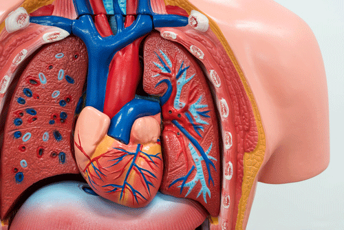
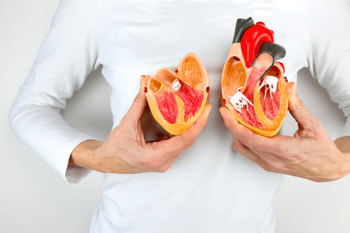 Ventricular diseases refer to an array of diseases that arise due to some serious defects in the ventricular structures, that are normally present from birth. A few commonly studied defects are- defects in the ventricular septum, ventricular valve dysfunctions, ventricular pumping abnormalities and problems detected in the ventricular walls. Any of these defects can cause a reverse (backward) flow of venous blood (de-oxygenated blood), which may get accumulated in different parts of the body and give rise to numerous systemic complications. One such complication is the formation of varicose veins in the lower extremities.
The next few sections will discuss the different types of ventricular diseases, their symptoms, and the possible systemic complications. The diagnosis and the treatment techniques will also be discussed in details.
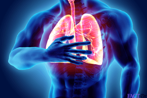
Ventricular hypertrophy
 It is pathological condition resulting from the thickening of the walls of the ventricles, that is, the lower chambers of the heart. It is mainly of three types- Right Ventricular Hypertrophy, Left Ventricular Hypertrophy, and Biventricular Hypertrophy. Right and left ventricular hypertrophy refers to the thickening of the ventricular walls of the right and the left ventricles respectively. Biventricular Hypertrophy refers to the thickening of the walls of both the ventricles.
There is another classification- Healthy and unhealthy cardiac hypertrophy. Healthy cardiac hypertrophy is basically the response to rigorous physical exercise or pregnancy. This condition leads to an increase in the mass of the heart muscles (especially the ventricular muscles) and an abnormal elevation in the pumping rate of the heart. Unhealthy cardiac hypertrophy manifests as a response to stress or certain diseases like hypertension, cardiac muscle injury, neurohormones or cardiac failure.
Ventricular tachycardia
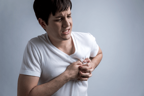
Ventricular tachycardia (VT) is a type of irregular and fast heart rate that arises from abnormal electrical activities in the ventricles of the heart. This usually occurs as transient episodes with some noticeable symptoms like palpitations, lightheadedness, chest pain and discomfort. Two main complications may arise in this case- ventricular fibrillation and cardiac arrest.
Ventricular tachycardia may result from coronary cardiac disease, aortic stenosis, electrolyte problems, cardiomyopathy or a past occurrence of heart attack. Based on morphology, ventricular tachycardia can be classified as-
- Monomorphic Ventricular Tachycardia: Here the appearance of all the beats match each other in each lead of a surface electrocardiogram. It is of two types- scar-related tachycardia and RVOT tachycardia.
- Polymorphic Ventricular Tachycardia: It shows beat-to-beat variations in morphology and appears as cyclical progressive changes in the cardiac axis.
Another classification is done based on the duration of episodes. These are-
- Non-sustained ventricular tachycardia (rhythm self-terminates within 30 seconds)
- Sustained ventricular tachycardia (rhythm lasts more than 30 seconds)
On the basis of the symptoms, VT can be loosely classified as – pulseless VT and VT with cardiac output.
Ventricular dyssynchrony
As the name implies, this condition is marked by a lack of synchrony or a difference in timing of the contractions of the different ventricles in the heart. If the differences are very large, cardiac output is reduced and this may even lead to cardiac failure.
Three different types of dyssynchrony may be observed-
- Atrioventricular dyssynchrony: In this case, there is a noticeable difference in timing between the auricular and the ventricular contractions.
- Interventricular dyssynchrony: In this case, there is a detectable time difference between the systolic contractions of the right and the left ventricles.
- Intraventricular dyssynchrony: Here, the timing in the sequence of activation and contractions of the different segments of the lower ventricular wall becomes abnormal.
In all the three types, the alterations in timing lead to certain abnormal behavior of the myocardial tissues. This often leads to mechanical dyssynchrony.
Ventricular aneurysm
It is one of the many systemic complications that are observed after a cardiac arrest. In this condition, a bulging or “pocketing” of the wall or the lining of a vessel at the basal portion of the septum or within the aorta occurs. The bulges mainly form due to the bubbling up of a weakened tissue, which then gets filled with blood. This can obstruct the passages that emerge from the heart, which in turn leads to a sluggish flow of blood to the different organs. The disease progresses at a slow pace and is often perceived as a pseudoaneurysm. The blood clots formed may get stuck within the blood vessels supplying to the limbs. This may result in ischemia and tissue death in the limbs.
Ventricular Outflow Tract Obstruction
It is a type of congenital defect where blockage of the right or left ventricular outflow tract is observed. This impedes the flow of blood from the ventricles and gives rise to a spectrum of disorders. It is of two types-
- Right ventricular outflow tract obstruction: It arises due to defects in the pulmonic valve, the supravalvar region, the infundibulum or the pulmonary artery.
- Left ventricular outflow tract obstruction: This occurs due to a defect in the aortic valve or a defect at the subvalvar or supravalvar level.
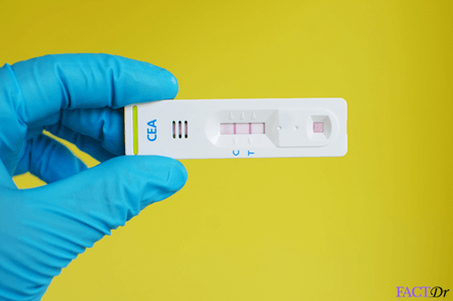
Diagnosis and treatment
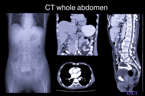
The main diagnostic tests are as follows-
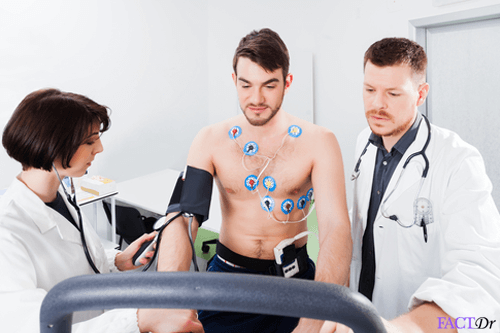
- Electrocardiogram
- Chest X-ray
- Echocardiogram
- Ultrasound
The left ventricular hypertrophy can be treated as follows:
Medications
Angiotensin-converting enzyme (ACE) inhibitors (Enalapril, captopril, lisinopril)- they help in widening the blood vessels and lowering the blood pressure so as to improve the blood flow to the heart.
Angiotensin II receptor blockers (ARBs) (losartan) – they have an effect similar to ACE inhibitors.
Beta-blockers (atenolol) – lowers the heart rate, reduces the blood pressure and protects the heart from the detrimental effects of stress hormones.
Calcium channel blockers (amlodipine, diltiazem) – these drugs also help in lowering the blood pressure
Diuretics (chlorthalidone, hydrochlorothiazide) – These medications facilitate blood flow and lower hypertension.
Surgical intervention- surgery may be required for aortic valve repair or replacement.
Sleep apnea can be treated by a machine that provides continuous positive airway pressure (CPAP) while sleeping. This helps in keeping your airways open and getting oxygen to maintain blood pressure levels.
Ventricular tachycardia (V-tach) can be treated as follows:
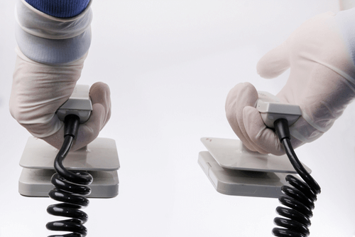
The normal heart rate is restored by means of a jolt of electricity via defibrillation or cardioversion. Defibrillation can be executed by means of an automated external defibrillator (AED). Cardioversion can be performed in a hospital via a cardioversion machine so as to monitor the heart rhythm before and after the shocks are delivered.
Prevention of episodes can be done by catheter ablation, anti-arrhythmic medications, open-heart surgery, and implantable cardioverter-defibrillator. Treating an underlying health condition is essential to prevent future episodes of V-tach. In general, prevention is mainly by following a healthy and balanced diet. Going for regular screenings and checkups is recommended so as to diagnose an underlying condition in early stages. Quitting tobacco consumption, smoking, drug abuse, excessive alcohol consumption and other detrimental habits is essential.
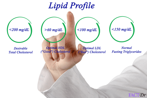
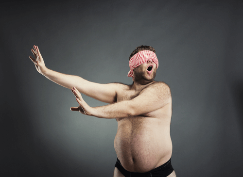
Physical activity is of vital importance to keep blood pressure, blood cholesterol and the total body weight under control.
- https://www.cdc.gov/heartdisease/facts.htm
- https://www.mayoclinic.org/diseases-conditions/ventricular-tachycardia/diagnosis-treatment/drc-20355144
- https://www.mayoclinic.org/diseases-conditions/ventricular-fibrillation/diagnosis-treatment/syc-20364524
- https://www.mayoclinic.org/diseases-conditions/left-ventricular-hypertrophy/diagnosis-treatment/drc-20374319
Dos and Don'ts
Dos
- Maintain a healthy lifestyle and diet. A low-sodium and low-fat diet is usually recommended.
- Go for a regular physical activity of at least 30 to 60 minutes.
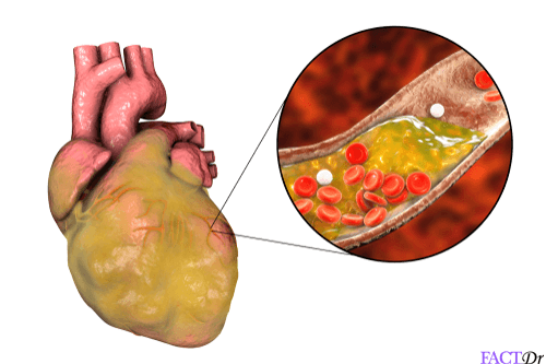
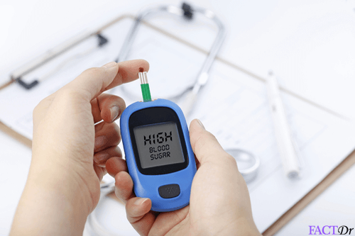
- Keep your blood pressure and cholesterol levels under check. Uncontrolled blood pressure and cholesterol levels are potential risk factors for ventricular diseases.
Don'ts
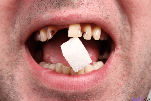
- Smoke tobacco. Smoking can multiply your risk of getting ventricular diseases.
- Have high alcohol intake. Drinking in moderation is advised otherwise it may have detrimental effects on the heart. Females and males above 65 years of age can have up to 1 drink in a day and males below the age of 65 years can have 2 drinks per day.
- Neglect your follow-up visits with the cardiologist. Follow-up care is crucial for recovery. The health can be monitored and necessary dose changes can be made.
Help Others Be Fit
Related Conditions


Trending Topics


About
Related Conditions

Subscribe to free FactDr newsletters.
REVAMP YOUR
LIFE
HEALTH
WELLNESS

If you're enjoying our website, we promise you'll absolutely love our new posts. Be the first one to get a copy!
Get factually correct, actionable tips delivered straight to your inbox once a week.
We hate spam too. We will never share your email address with anyone. If you change your mind later, you can unsubscribe with just one click
By clicking Subscribe, I agree to the FactDr Terms & Conditions & Privacy Policy and understand that I may opt out of FactDr subscriptions at any time.
×
How can we improve it?
×
Happy to know you loved our article!
Did it give you information that you used / can use in your life?