
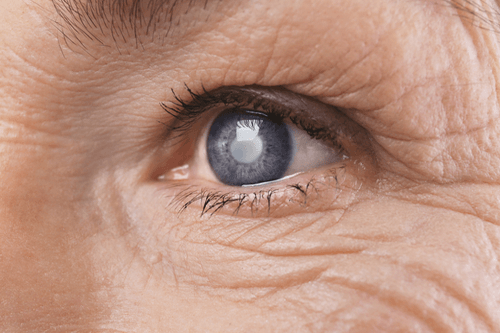
Glaucoma
Last Updated December 20th, 2021
Glaucoma and vision loss
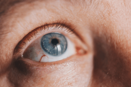
A gradual build-up of pressure on the eye which damages the optic nerve and leads to blindness is termed as Glaucoma. This progressive disease is characterized by a slight peripheral vision loss initially to complete blindness if left untreated. The optic nerve is the nerve that transmits optical nerve impulses from retina to the brain which makes vision possible. When this nerve gets pressurized (intraocular pressure), the power of vision is considerably affected leading to glaucoma. The vision loss may also occur due to the poor blood supply to this nerve. The onset of this condition doesn’t cause sudden blindness. The loss of vision is a gradual process and it may even take years for complete blindness to occur yet it is identified as one of the leading factors causing irreversible blindness.
Glaucoma mainly exists in two types – Open-Angle Glaucoma and Angle-Closure Glaucoma. Majority of glaucoma cases fall in the open-angle glaucoma category. A clear fluid flows through the anterior chamber of the eye nourishing the tissues. At the point where the iris meets the cornea, there is a spongy network like a drain through which this liquid flows. When the flow of this fluid is too slow through the drain, it mostly deposits along the drain causing a pressure build-up in the eye. This pressure causes optic nerve damage and leads to open-angle glaucoma. In case of Angle-closure glaucoma, the angle between the iris and cornea is too small for the fluid to drain properly leading to pressure build-up by the fluid.
Causes and risk factors
As explained above, Glaucoma is a result of optic nerve damage caused by pressure build-up in the eye.
This pressure is created either due to a small angle between iris and cornea (angle-closure) or due to certain factors which slow down the liquid flow in the drain (open-angle).
There are a number of reasons which could be causing the block in the flow of the liquid through the eye such as:
- Physical injury to the eye.
- Exposure to certain chemicals may create a blockade in liquid flow
- Eye infections which block blood vessels of the eyes.
- In some people, increased caffeine intake has resulted in intraocular pressure.
These factors have led to glaucoma in a majority of people yet the presence of these factors doesn’t necessarily mean that the individual will suffer from glaucoma. In addition to these, there are certain risk factors associated with this condition:
- Being 40 years or above.
- Having a family history of glaucoma.
- Belonging to certain ethnicities such as Japanese, Hispanic, African-American, Irish or Scandinavian.
- Prolonged exposure to certain steroids.
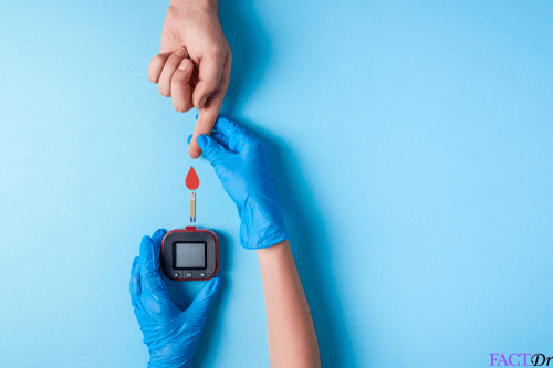
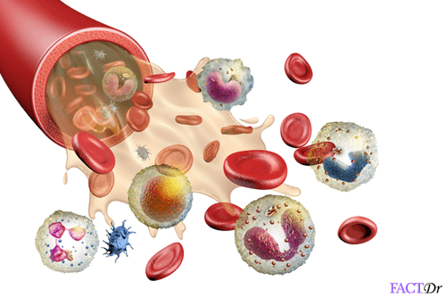
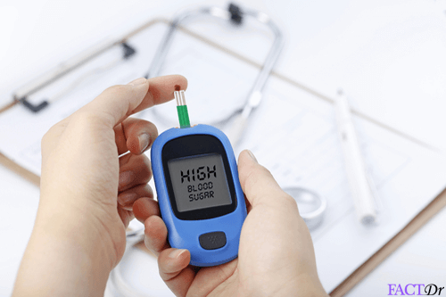
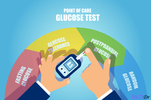
- Having diabetes which causes a poor blood flow to the eyes.
- Thinner cornea
- Myopia or hypermetropia
Signs and symptoms
 The characteristic trademark of this condition is the onset of peripheral or side vision loss which is not readily detected in the initial stage.
Very few individuals experience hazy or blurred vision and can see “halo-like” structures surrounding light sources.
Since the symptoms go undetected in the early stages, this condition is also called as the “sneak thief of vision”.
In many cases, when the condition manifests in the acute form, the onset of symptoms are more dramatic.
Acute glaucoma is identified by a severe headache and pain in the eyes followed by nausea. Visual blurring is more pronounced in this case.The pupil remains in the position of mid-dilation and may also turn oval in shape. Redness in the eye prevails with the cornea appearing cloudy. In both the cases, the individual starts experiencing tunnel vision.
What are the different types of glaucoma?
The classification of the disease is done based on the presence of medical conditions that may cause an increase in intraocular pressure. The different subtypes will be discussed in separate sections.
Open-angle glaucoma
This is a major retinal disorder. It is the most widespread form of glaucoma (occurs in 90% of the cases). It occurs due to progressive clogging of the drainage paths in the eye chamber, which leads to an increased intraocular pressure. In this condition, the eye fluid travels drain very slowly via the connecting meshwork between the cornea and the iris. In due course of time, this leads to the damage of the optic nerve. Loss of vision occurs in the late stages.
Types of open-angle glaucoma
Open-angle glaucoma has three subtypes-
Primary open angle glaucoma: This type is common in people who are over 50 years old. The cornea gradually adapts to the increase in eye pressure but does not swell. The functioning of the trabecular meshwork (a group of tissues present at the base of the cornea) is normal in this case. In old people, the lesser number of cells is present in this meshwork which may lead to glaucomatous symptoms. A faulty drainage system or some enzymatic abnormalities may also contribute to the disease.
- Secondary open angle glaucoma: This disease occurs due to the presence of any kind of substance (red blood cells or pigment exfoliation matter) that blocks the flow of intraocular fluid through the trabecular meshwork. This condition, along with some other conditions like inflammation, ischemia and trauma can lead to a high intraocular pressure and glaucomatous symptoms.
- Pigmentary glaucoma: This is an inherited retinal disease. Here the iris pigments shed and accumulate in the aqueous humor. This leads to the clogging of the pores of the trabecular meshwork, thereby increasing the pressure.
Normal tension glaucoma
In this case, the intraocular pressure remains more or less normal. Optic nerve damage and loss of vision are the main features of this type of retinal disorders. This is attributable to the damage of the cells that transmit impulses from the retina to the brain, which causes a poor blood flow condition.
Closed-angle glaucoma
In closed-angle glaucoma, the gap between the cornea and the iris is shortened, which causes an obstruction in fluid flow. This happens due to a smaller size of the anterior chamber. The angle at the junction of the cornea and the iris becomes smaller than 45o in these patients, which hinders the normal flow of eye-fluid and raises the eye-pressure.
Types of closed angle glaucoma
This disease has the following subtypes-
- Acute glaucoma: In this type, the intraocular pressure increases abruptly, within a few hours. This causes redness and cloudiness of the cornea and leads to blurred vision.
- Chronic closed-angle glaucoma: This disease develops gradually over a number of years. It leads to an increase in pressure and a gradual of vision.
Congenital glaucoma
This type of childhood glaucomatous disease is usually present at birth. It occurs due to some basic structural defects in the drainage system of the eyes.
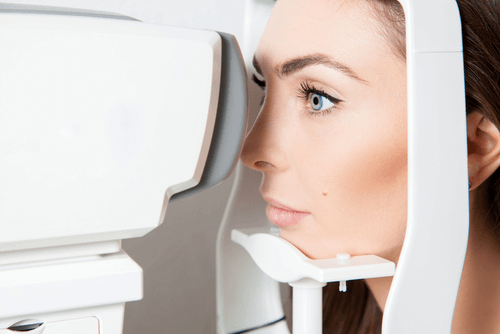
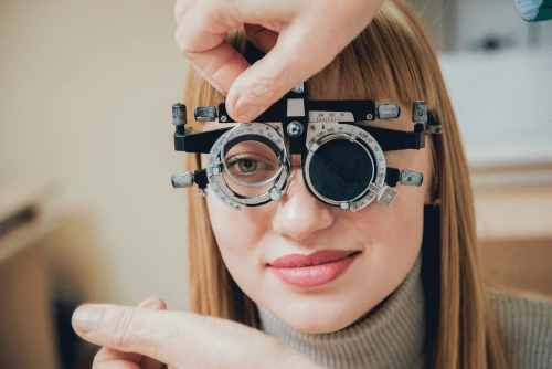
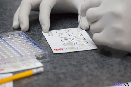
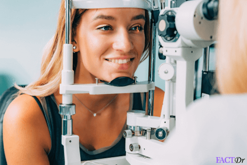
Diagnosing glaucoma
There are a series of comprehensive eye tests through which the type and degree of glaucoma occurrence can be diagnosed clearly:
- Tonometry which calculates the intraocular pressure and pachymetry which determines the corneal thickness. These tests give a clear idea about any abnormalities within the anatomy of the eye. Both of these tests are conducted by adding numbing drops to the eyes.
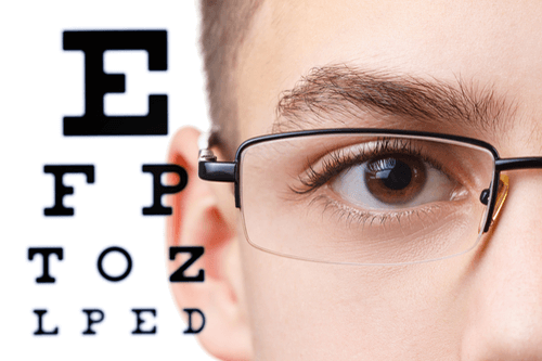
- Visual Acuity Test will be conducted by your ophthalmologist to determine the quality of your vision for different distances. Similarly, a Visual Field Test is conducted to find out of the person is suffering from peripheral vision loss.
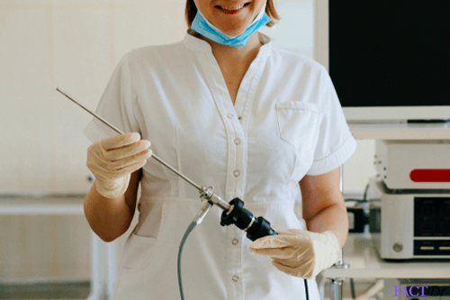
- Dilated Eye Test or Ophthalmoscopy is also conducted to check for the shape and the coloration of the optic nerve. In this method, eye drops are used to dilate the eye pupil and then through a magnifying device, the doctor observes the optic nerve structure to detect any damage.
- The optic nerve is a collection of more than a million nerve fibers. To detect any anomaly in this, the ophthalmologist may also additionally conduct a Nerve Fibre Analysis to examine the optic nerve thickness.
Treating glaucoma
 The blindness caused by glaucoma is permanent hence early detection of this condition is imperative for the treatment to be successful.
The aim of every treatment methodology is to control and decrease the intraocular pressure which can be achieved through the following methods:
- Medications – The medications prescribed for glaucoma patients are usually eye-drops and pills which lower the build-up of eye pressure. These either decrease the liquid flow in the eye or facilitate easy draining of the fluid. A strict adherence to the medication protocol is important here. These medications may lead to certain side-effects such as Hyperpigmentation of the eye and growth of eyelashes.
- Laser Trabeculoplasty – In this method, laser beams are used to reconfigure the drain system in your eye to enable the liquid to flow smoothly within the eye. Glaucoma medications are also prescribed in addition to this procedure. Regular follow-up is important as the laser procedure may cause inflammation in the eye in some patients.
- Glaucoma Surgery – Certain conventional surgery methods can also be conducted to treat glaucoma, especially when it is congenital in nature. Various surgical methods include Canaloplasty, trabeculectomy, glaucoma drainage implants, and sclerectomy (non-penetrative). Surgeries are 60%-80% effective in completely curing this disease.
Preventive measures
While complete preventive measures for glaucoma don’t exist since this condition is caused by a host of factors and in many cases, is congenital. Early detection and proper treatment is the most effective solution for glaucoma. Regular eye check-ups should be taken up by people who have crossed their 40s. Injury to the eye should be immediately taken care of. Wearing protective head-gear during sports, swimming, or any hazardous activity may reduce the chances of physical damage to the eye.

Display this infographic on your website
Dos and Don'ts
Dos
- If you are taking eye-drops for glaucoma, make sure that you are following the exact time-schedule provided by your doctor.
- Provide a complete list of your medications to your eye doctor since glaucoma medications may react with certain drugs.
- If you have not been diagnosed with glaucoma, it is still wise to get regular eye check-ups after the age of 40.
Don'ts
- Expose your eyes to TVs, computer screens, phones, etc for longer durations.
- Since there is a loss of peripheral vision, remove carpets and rugs from the floor to avoid the patient from tripping over. Add colored handrails for marking stairways and handrails.
Help Others Be Fit
Related Conditions

Trending Topics


About
Related Conditions


Subscribe to free FactDr newsletters.
REVAMP YOUR
LIFE
HEALTH
WELLNESS

If you're enjoying our website, we promise you'll absolutely love our new posts. Be the first one to get a copy!
Get factually correct, actionable tips delivered straight to your inbox once a week.
We hate spam too. We will never share your email address with anyone. If you change your mind later, you can unsubscribe with just one click
By clicking Subscribe, I agree to the FactDr Terms & Conditions & Privacy Policy and understand that I may opt out of FactDr subscriptions at any time.
×
How can we improve it?
×
Happy to know you loved our article!
Did it give you information that you used / can use in your life?